Like
Share
멍실장
강아지 종양 CT·MRI 스테이징 증상과 원인, 진단·치료와 관리 포인트 총정리

Complete Guide to CT and MRI Scans for Canine Tumors — From Staging to Treatment Planning

Oncology/CancerDiseasesMeongsiljang Veterinary Advisory Board

CT and MRI scans are essential for accurately staging tumors in dogs. This article provides a comprehensive guide covering causes, symptoms, diagnostic methods, treatment strategies, and key care considerations.

What Is CT and MRI Staging for Canine Tumors?

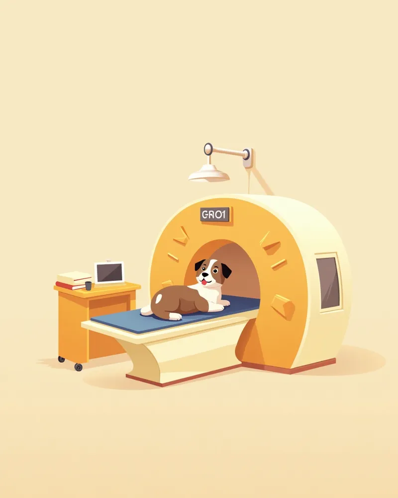
A dog undergoing a CT scan, with a tumor visible in the abdomen.
CT and MRI staging for canine tumors are imaging techniques used to precisely determine the tumor’s location, size, and extent of invasion. These tests are essential for developing a treatment plan.
CT scans: Clearly highlight differences in density between bone and soft tissue, making them ideal for assessing bone involvement.
MRI imaging: Provides high-resolution views of soft tissue anatomy, making it optimal for diagnosing brain and spinal cord tumors.
Accurate staging directly influences decisions regarding surgical feasibility, radiation therapy planning, and the assessment of metastasis.

Main Causes and Risk Factors for Canine Tumors

Dogs face a lifetime risk of cancer similar to that of humans. However, since most cancers arise randomly, it is often difficult to pinpoint a single cause. Therefore, early detection and management are more important than trying to determine the exact cause.
Breed Predisposition: Certain breeds are reported to have a higher tendency to develop specific tumors. For example, herding dogs and Golden Retrievers are known to be predisposed to certain oral tumors.
Age: Tumors are more commonly diagnosed in middle-aged to senior dogs, with some types typically appearing around 7 to 10 years of age.
Chronic Lesions: Areas with long-standing or non-healing lesions can undergo changes, so it is advisable to have them checked through examinations.
Immune Status: It is also helpful to assess overall health and immune status.
Since it is difficult to clearly identify the cause, regular observation and routine check-ups for early detection are the most important steps.
Suspected tumor on your dog’s skin: genetic, environmental, and aging factors

Key Symptoms Observed in Canine Tumor Staging

The symptoms of tumors in dogs vary depending on their location and type. Careful observation is essential for early detection.
Weight loss: Unexplained weight loss may occur due to a decreased appetite or metabolic changes.
Persistent pain: The dog may lick specific areas or show restricted movement.
Swelling or tumor masses: Lumps may develop under the skin or internally.
Behavioral changes: Signs include lethargy, excessive sleepiness, reduced concentration, or increased aggression.
Difficulty breathing: Tumors in the lungs or chest cavity can cause rapid or labored breathing.
If these signs persist, consult your veterinarian immediately.
Lethargic dog with weight loss, symptom icon displayed

Emergency Signs Requiring Immediate Veterinary Visit

If your dog shows any of the following symptoms, take them to the vet immediately. In particular, with brain or spinal cord tumors, quick action can significantly impact survival rates.

CT and MRI Scan Procedures for Diagnosing Canine Tumors

For accurate tumor staging, CT and MRI scans are essential diagnostic tools recommended by veterinarians, each with its own advantages.
CT Scan: Provides rapid imaging of bone and organ structures, making it particularly useful for determining whether a tumor has invaded the bone.
MRI Scan: Delivers detailed visualization of subtle changes in soft tissues such as the brain, spinal cord, and muscles, making it ideal for assessing tumor boundaries and the extent of invasion into surrounding tissues.
These scans are performed under anesthesia, and the veterinarian will determine the most appropriate imaging method based on the patient’s condition.
A veterinarian reviewing a puppy’s brain MRI scan

Treatment Methods and Step-by-Step Approach for Canine Tumors
Treatment for canine tumors varies depending on the tumor type, location, and staging results, and, much like cancer treatment in humans, it involves a comprehensive approach using surgery, medication, and radiation therapy.
Surgical treatment: When a tumor is localized and removable, surgery is the primary option.
Radiation therapy: This is used either as a standalone treatment or as an adjuvant therapy after surgery for tumors that are sensitive to radiation, such as mast cell tumors and melanomas.
Chemotherapy: This is used for tumors that have spread throughout the body, such as lymphoma, or to suppress distant metastasis after the removal of malignant tumors.
Immunotherapy: Approaches like the melanoma vaccine are being explored, but because their efficacy has not yet been sufficiently proven, they are carefully considered only as adjunctive treatments.
If metastasis has already progressed, treatment may focus on palliative care to alleviate pain. The treatment plan is determined in consultation with your veterinarian based on a comprehensive assessment of the tumor’s status.
A puppy receiving radiation therapy at a veterinary clinic

Home Tumor Management and Care Points

During treatment, home care plays a vital role in improving your dog’s quality of life.
Dietary management: Provide a nutritionally balanced diet to help maintain immune function.
Pain management: Administer pain medication exactly as prescribed by your veterinarian.
Activity management: Avoid excessive exercise and maintain a comfortable environment.
Regular check-ups: Continue periodic CT and MRI scans after treatment to monitor for recurrence.
Emotional well-being: Your warm attention and a stable daily routine support your dog’s recovery.
A dog resting comfortably next to its food bowl.

Breed-Specific Precautions and Tips for Preventing Recurrence

Certain breeds are more susceptible to specific tumors, making prevention and early detection crucial. Senior dogs, in particular, should undergo regular veterinary check-ups.

Reviewed by a veterinarian

Dr. Tony — Punnawat Phongkittirak

Dr. Tony — Punnawat Phongkittirak

Veterinarian

A veterinarian who majored in veterinary medicine at Khon Kaen University, Thailand, and completed the IVSA program at North Carolina State University in the United States. Drawing on clinical experience at animal hospitals, he works in the pet healthcare field and is dedicated to building a digital care environment that connects pet parents with veterinarians.

Frequently Asked Questions

Which is more accurate, CT or MRI?
CT scans provide clear images of bone and organ structures, while MRI offers more detailed visualization of soft tissue. The appropriate imaging test is selected based on the tumor's location.
How often should tumor staging tests be performed?
Before starting treatment, it’s important to determine the stage of the disease, and after treatment, regular follow-up exams should be conducted as recommended by your veterinarian. Consistent monitoring is essential to check for recurrence or metastasis.
Can CT and MRI scans be performed without anesthesia?
If your dog moves during the imaging, the resulting blurriness can make diagnosis difficult. Therefore, to ensure clear and accurate images, the procedure is typically performed under anesthesia or sedation. Your veterinarian will determine the most appropriate approach based on your pet's individual condition.
Does a tumor always require surgical removal?
Not all tumors are candidates for surgery. We determine the treatment plan by carefully evaluating the tumor’s location, size, and whether it has spread.
Is chemotherapy scary for dogs?
Side effects can vary from pet to pet. Temporary loss of appetite and fatigue may occur, but your veterinarian will manage any side effects throughout treatment, so close monitoring is essential.

CT vs MRI: Comparing Differences in Tumor Staging

ItemCTMRI
Main advantagePrecise imaging of bone structuresHigh-resolution images of soft tissue
Suitable tumor typesBone tumors, lung tumorsBrain tumors, spinal cord tumors
Examination timeAbout 15–30 minutesAbout 45–60 minutes
Anesthesia requiredRequiredRequired

The appropriate imaging test must be selected according to the tumor's location and characteristics to enable an accurate diagnosis.

Share

Related Guides

References

[1] Clinical Medicine of the Dog and Cat, 4th Ed. Chapter 14: Disorders of the Nervous System and Muscle, 2023

[2] Advanced Monitoring for Small Animal Emergency and Critical Care, 2nd Ed. 2022

[3] Veterinary Neuroimaging: Principles and Practice. 2021

This information is based on veterinary literature and does not replace diagnosis or treatment. Please consult a veterinarian for specific health concerns.

Metapet Co., Ltd. | CEO: Park Sung-yong | Business Reg. No. 417-88-02562 | Seoul, South Korea | Customer Center

Dog Tumor CT & MRI Guide: From Staging to Treatment Plans | Meongsiljang