Like
Share
멍실장
강아지 동공 이상(이방동공 등) 증상과 원인, 진단·치료와 관리 포인트 총정리

If Your Dog’s Pupils Are Unequal in Size — Causes of Anisocoria and When to Visit the Vet

Eye HealthDiseasesMeongsiljang Veterinary Advisory Board

Abnormalities in a dog’s pupils, such as anisocoria (unequal pupil size), occur when one pupil differs from the other in size or light response and can signal neurological issues or eye disease. Early detection and accurate diagnosis are essential.

What Are Pupillary Abnormalities in Dogs (Such as Anisocoria)?
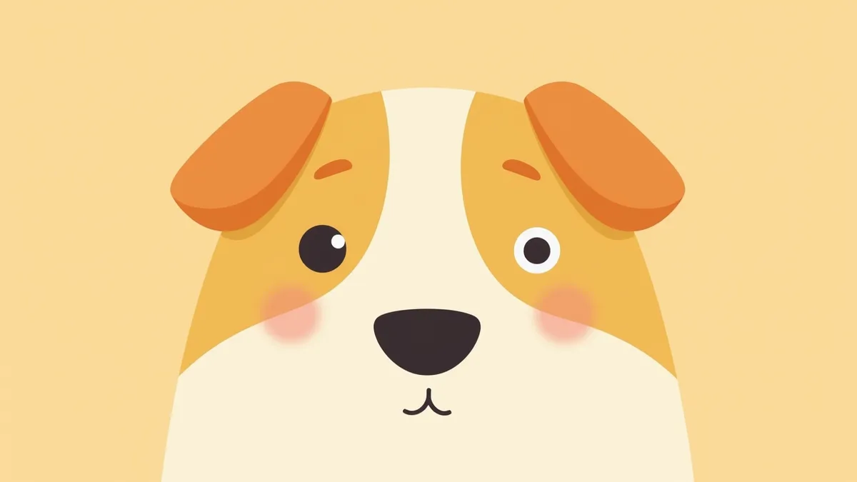
A dog’s eyes with one pupil dilated and the other constricted
Abnormalities in a dog’s pupils, particularly anisocoria, refer to a condition where one pupil differs from the other in size or light response. This may present as one pupil becoming larger (mydriasis) or smaller (miosis). Such abnormalities can arise from a variety of causes, ranging from ocular and periocular issues—such as sympathetic nerve damage leading to Horner’s syndrome, glaucoma, uveitis, optic neuritis, and retinal diseases—to systemic conditions like hypertension. Immediate veterinary attention is required if a pupil suddenly dilates or constricts. In particular, a lack of response to light or accompanying visual disturbances may signal neurological or retinal problems. Early detection and accurate diagnosis are key to effective treatment. Because pupillary abnormalities can reflect overall systemic health beyond just eye issues, they warrant careful attention.

What Are the Main Causes of Anisocoria?
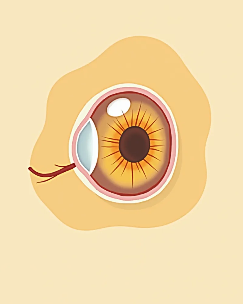
Anisocoria, or unequal pupil sizes, can stem from a wide variety of causes. Damage to the sympathetic nerves supplying the eye can lead to Horner’s syndrome, which causes one pupil to constrict (miosis); this is one of the more common causes of anisocoria. Horner’s syndrome is most frequently idiopathic, resulting from postganglionic nerve lesions of unknown origin, but it can also be caused by mediastinal tumors, brachial plexus injuries, or otitis media. Additionally, conditions affecting the eye itself or the entire body—such as closed-angle glaucoma, anterior uveitis, optic neuritis, retinal blindness, iris atrophy, systemic hypertension, and aqueous misdirection syndrome—can also cause a difference in pupil size between the two eyes. Because accurate diagnosis is essential for effective treatment, thorough examination is crucial. A comprehensive evaluation is necessary, as multiple factors may often be involved simultaneously.
Anatomy of the nerves and muscles in a dog's eye

Main Symptoms and Signs

The primary sign of pupillary abnormalities in dogs is when one pupil is larger or smaller than the other. The pupil may fail to constrict when exposed to light, or vision disturbances may occur simultaneously. In Horner’s syndrome, caused by damage to the sympathetic nerves supplying the eye, miosis (constricted pupil) is accompanied by ptosis (drooping of the upper eyelid), protrusion of the third eyelid (nictitating membrane), and enophthalmos (sunken eye). If the eye is painful, blepharospasm (frequent blinking) and excessive tearing may also appear. These signals indicate that immediate veterinary care is necessary.
Anisocoria: A condition in which the two pupils appear to be different sizes.
Absent light reflex: When the pupil does not constrict upon exposure to light.
Signs of Horner’s syndrome: Occurs when miosis, ptosis, protrusion of the third eyelid, and enophthalmos are present together.
Visual disturbances: Includes inability to see surroundings or a diminished menace response.
Epiphora (excessive tearing): Tearing that flows around the eye, often indicating ocular pain.
A dog’s eyes with pupils of different sizes

When Immediate Veterinary Care Is Needed

If your pet’s pupils suddenly dilate or constrict, their vision becomes blurry, or they no longer react to light, seek veterinary care immediately. These signs may indicate conditions requiring prompt treatment, such as Horner’s syndrome (nerve damage), glaucoma, optic neuritis, or systemic hypertension.

Diagnostic Tests for Accurate Diagnosis
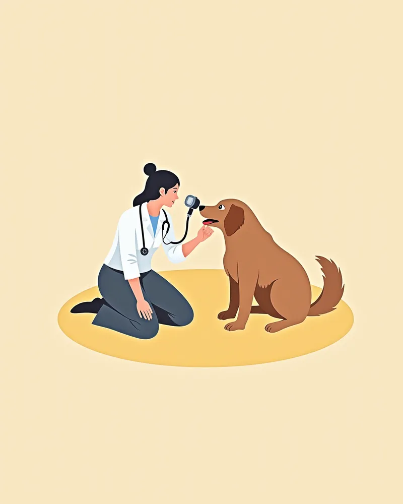
To accurately diagnose abnormal pupil dilation in dogs, multiple tests are typically performed together. First, a neuro-ophthalmic exam assesses the pupillary light reflex (direct and indirect PLR), menace response, dazzle reflex, and palpebral reflex, while comparing both pupils using reverse illumination, where light is shone at each eye individually. Brain imaging (CT or MRI) is crucial for detecting nerve damage, tumors, or lesions in the brain and meninges. Blood tests (CBC and chemistry panels), infectious antibody tests, and cerebrospinal fluid analysis help evaluate inflammation and infection while ruling out systemic diseases. Electroretinography may also be used to differentiate retinal diseases. Since effective treatment is difficult without identifying the exact cause, a comprehensive diagnostic approach is essential.
A veterinarian examining a puppy's eyes with an ophthalmoscope

Treatment Methods and Step-by-Step Management
Treatment depends on the underlying cause. If inflammation is the culprit, we use local or systemic anti-inflammatory medications (steroidal or non-steroidal) along with immunomodulators. If glaucoma is present, we administer medications to lower intraocular pressure, such as carbonic anhydrase inhibitors, or miotics to constrict the pupil. In cases where surgery is necessary—such as for tumors, trauma, or advanced glaucoma that cannot be controlled with medication—we proceed with surgical intervention. Even after addressing the root cause, long-term management is often required to prevent recurrence or secondary glaucoma, making ongoing veterinary monitoring essential.
Medication Therapy: Anti-inflammatory drugs and immunomodulators are prescribed for inflammation and infection, while intraocular pressure-lowering agents are used if pressure is elevated.
Surgical Treatment: Surgery may be necessary for tumors, trauma, or glaucoma that cannot be managed with medication alone.
Ongoing Medication Management: In many cases, long-term treatment is needed to maintain comfort.
Regular Check-ups: Monitoring is required to watch for recurrence and secondary glaucoma, even after the underlying cause has been resolved.
A veterinarian administering eye drops to a puppy

Home Care and Precautions

At home, regularly monitor your dog’s pupil changes and check for light reflexes. It’s important to minimize stress and maintain a calm, stable environment. When administering medication, give it at consistent times and adjust only as directed by your veterinarian. Contact us immediately if you notice signs of anxiety or unusual behavior. Maintain a quiet environment: Reduce external stimuli to help lower stress. Regular monitoring: Check pupil size and response daily. Consistent medication schedule: Administer medication at the same time each day. Stay in touch with your vet: Contact us right away if any abnormal symptoms appear.
A dog resting comfortably in a quiet environment.

Breed-Specific Considerations and Prevention of Recurrence

Certain breeds are more prone to eye conditions. For example, Collies may develop Collie Anomaly, Golden Retrievers are susceptible to uveitis, and Pugs often experience pigmentary keratitis. These conditions can lead to pupil abnormalities, so regular eye exams and minimizing external irritants are essential to prevent recurrence.

Reviewed by a veterinarian

Dr. Tony — Punnawat Phongkittirak

Dr. Tony — Punnawat Phongkittirak

Veterinarian

A veterinarian who majored in veterinary medicine at Khon Kaen University, Thailand, and completed the IVSA program at North Carolina State University in the United States. Drawing on clinical experience at animal hospitals, he works in the pet healthcare field and is dedicated to building a digital care environment that connects pet parents with veterinarians.

Frequently Asked Questions

My dog’s pupils suddenly dilated. Is this dangerous?
Yes, sudden dilation or constriction of the pupils can be a sign of serious conditions such as nerve damage, glaucoma, optic neuritis, or systemic hypertension. Immediate veterinary care is necessary.
Can anisocoria be treated?
It depends on the underlying cause. While some conditions, such as inflammation, can be effectively managed with medication or surgery, others like glaucoma may result in irreversible vision loss. Accurate diagnosis and targeted treatment are essential, and in many cases, ongoing care is necessary.
Can long-term anisocoria affect vision?
It depends on the underlying cause. Persistent nerve damage or glaucoma can lead to vision loss. Early diagnosis and treatment are crucial.
How can you check for abnormal pupil reactions at home?
In a dark room, shine a flashlight into each eye one at a time and compare the size of both pupils and their response to light. If you notice any differences, consult your veterinarian.
Can anisocoria be genetic?
In some breeds, genetic factors may play a role. For example, Collie eye anomaly is a breed-specific eye condition linked to genetic predisposition, so extra caution is advised for affected breeds.

Share

Related Guides

References

[1] Plumb's Veterinary Drug Handbook, 9th Edition, 2022

[2] Veterinary Ophthalmology, 5th Edition, 2020

[3] Small Animal Internal Medicine, 5th Edition, 2019

This information is based on veterinary literature and does not replace diagnosis or treatment. Please consult a veterinarian for specific health concerns.

Metapet Co., Ltd. | CEO: Park Sung-yong | Business Reg. No. 417-88-02562 | Seoul, South Korea | Customer Center

Unequal Pupil Size in Dogs: Causes of Anisocoria & When to See a Vet | Meongsiljang