Like
Share
멍실장
강아지 녹내장 — 급성 안압 상승 응급 신호와 치료

If Your Dog’s Eye Suddenly Swells and Becomes Red — Emergency Sign of Acute Intraocular Pressure Spike from Glaucoma

Eye HealthDiseasesMeongsiljang Veterinary Advisory Board

Canine glaucoma is an emergency eye condition in which abnormally high intraocular pressure damages the optic nerve. The golden window for preserving vision is within 24 to 48 hours after detection.

What Is Canine Glaucoma?
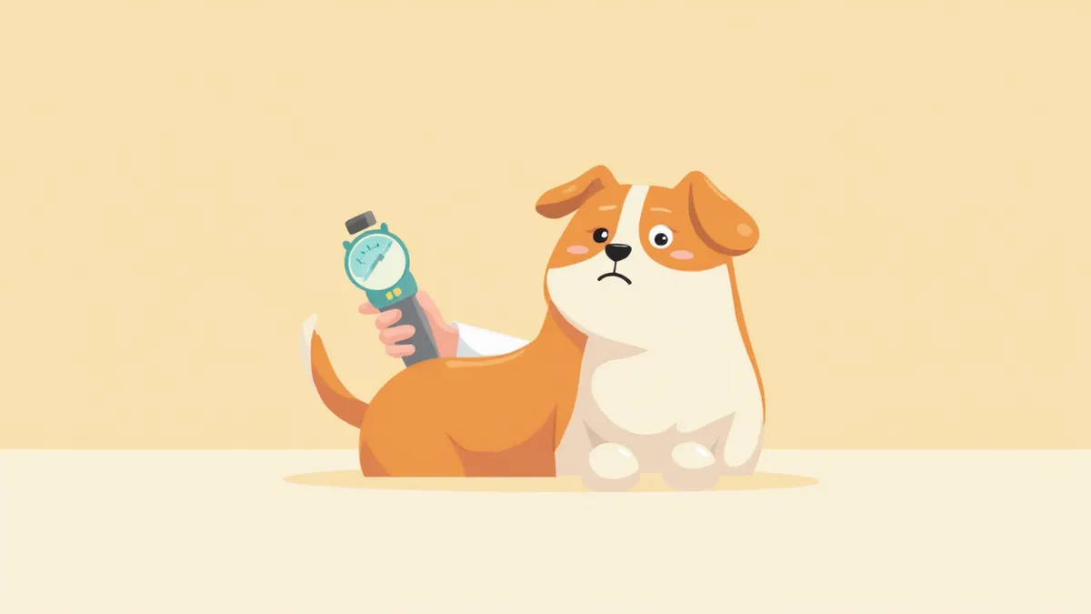
Emergency Guide for Canine Glaucoma
Canine glaucoma is an emergency eye condition in which abnormally high intraocular pressure damages the optic nerve. Primary glaucoma, in particular, often presents with a sudden spike in pressure—sometimes within just a few hours—so it’s crucial to seek veterinary care as soon as symptoms appear. If one eye becomes suddenly red, your dog struggles to keep it open due to pain (squinting), or the cornea turns cloudy, head to a 24-hour emergency animal hospital immediately, even at night. Once the optic nerve is damaged, it’s difficult to restore, and the longer the pressure remains elevated, the less likely vision can be recovered.

Causes — Primary and Secondary Types

Glaucoma is broadly categorized into two types based on its underlying cause. Primary glaucoma occurs when the drainage pathway for aqueous humor (the trabecular meshwork) is genetically narrow or blocked. Breeds prone to this condition include the American Cocker Spaniel, Basset Hound, Shih Tzu, and Shar-Pei. Secondary glaucoma develops when other eye diseases—such as uveitis, cataracts, lens luxation, intraocular hemorrhage, or tumors—obstruct the flow of aqueous humor. Secondary glaucoma is also quite common in dogs.

Checklist of Main Symptoms

If you notice any of the following signs, glaucoma should be suspected. However, these symptoms can also appear in other eye conditions such as uveitis, conjunctivitis, and keratitis, so a definitive diagnosis of glaucoma cannot be made based on symptoms alone. If you observe any of these signs, it is essential to have your pet’s intraocular pressure measured at a veterinary clinic using a tonometer for confirmation. - Eye redness: Prominent, dilated blood vessels in the white of the eye - Corneal clouding: The cornea appears bluish or hazy - Pupil changes: Reduced response to light, with the pupil becoming dilated (or, less commonly, constricted) - Increased tearing and discharge: Secretions resulting from pain - Eye rubbing: Rubbing the face with the front paws or against the floor - Decreased activity: Loss of appetite and hiding curled up in a corner
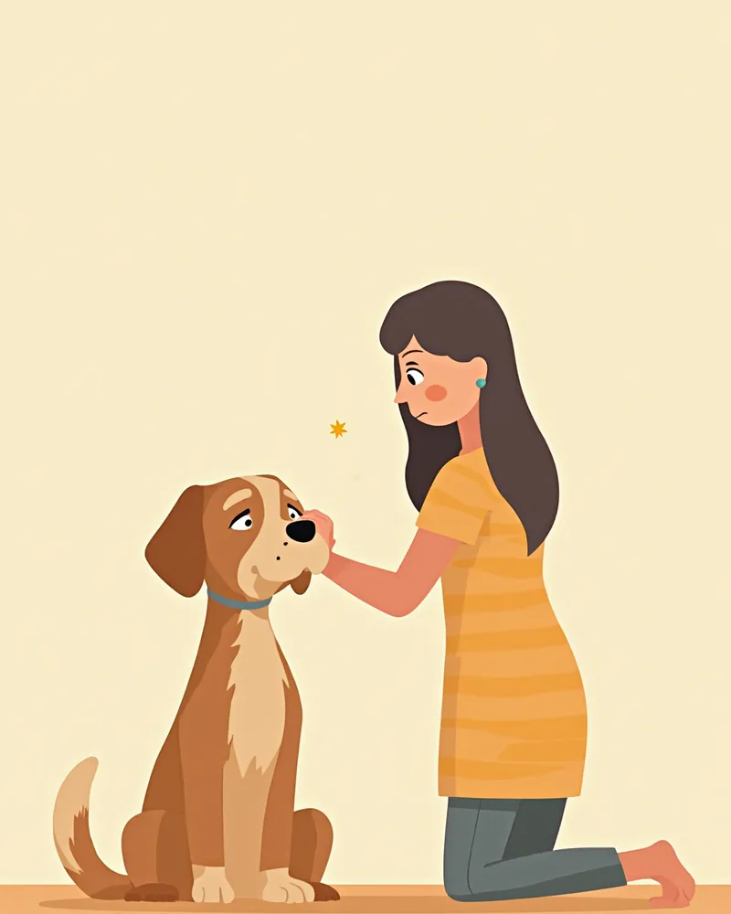
A pet parent checking for signs of glaucoma

Signs Requiring Immediate Visit to a 24-Hour Emergency Hospital

If any of the following symptoms are present, visit a 24-hour emergency animal hospital immediately, even in the middle of the night. Prolonged, significantly elevated intraocular pressure can rapidly damage the optic nerve and lead to blindness. - Sudden swelling or bulging of one eye (proptosis) - Sudden bluish or whitish discoloration of the cornea - Inability to open the eye at all, with vocalization due to pain - Marked difference in pupil size between the two eyes, or one pupil that is fixed and dilated

Diagnostic Tests Performed at the Veterinary Hospital

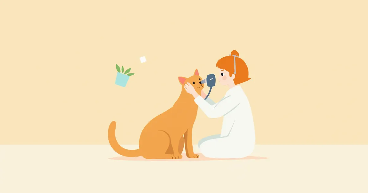
The cornerstone of glaucoma diagnosis is measuring intraocular pressure using a tonometer. If the pressure inside the eye is found to be higher than the normal range, glaucoma is suspected. Because external signs like redness or corneal clouding can overlap with other eye conditions, directly measuring the intraocular pressure is the most reliable way to confirm the diagnosis. Additionally, we perform fundus examinations to check for damage to the optic nerve head, and use gonioscopy or ultrasound to assess structural abnormalities in the aqueous humor drainage pathways. It is standard practice to examine both eyes, as primary glaucoma has a genetic predisposition, meaning that if one eye is affected, the other is at high risk of developing the condition as well.

Treatment — Step-by-Step Approach with Medication and Surgery
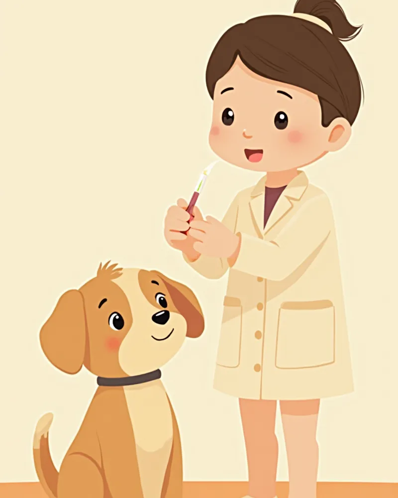
The primary goal of treatment is to rapidly lower intraocular pressure to protect the optic nerve.
Medical treatment: Prostaglandin analog eye drops (such as travoprost) are used to increase aqueous humor outflow, while carbonic anhydrase inhibitors reduce aqueous humor production. In emergency cases, intravenous mannitol may also be administered.
Surgical treatment: If intraocular pressure cannot be controlled with medication or if preserving vision is difficult, options include laser cyclophotocoagulation, aqueous humor drainage implants, or, in cases of vision loss, enucleation (eye removal). The exact dosage schedule and amounts are determined by a veterinary ophthalmologist.
Administering glaucoma eye drops

Comparison of Main Intraocular Pressure-Lowering Medications

ItemProstaglandin analogsCarbonic anhydrase inhibitorsBeta-blockers
Mechanism of actionIncreases aqueous humor outflowDecreases aqueous humor productionDecreases aqueous humor production
Onset of effectPer veterinarian's guidancePer veterinarian's guidancePer veterinarian's guidance
Primary timing of useEmergency & long-term managementLong-term managementAdjunctive use
Use in catsVeterinary judgment requiredVeterinary judgment requiredVeterinary judgment required

The onset time of each drug and its applicability to cats vary by individual and by agent. Always follow a veterinary ophthalmologist's prescription for specific drug selection and dosing schedules.

Key Points for Home Care

Here are the key points to keep in mind for home care after treatment: - Strict adherence to eye drop schedules: Missing the scheduled time for intraocular pressure (IOP) control medications can cause IOP to rise again. Setting an alarm is essential. - Use a harness instead of a collar: Pressure on the neck can affect IOP, so we recommend using a chest harness. - Avoid sudden excitement: Please minimize situations that cause excessive excitement or straining. - Regular check-ups for the other eye: Primary glaucoma has a genetic predisposition, and it often develops in the other eye over time. Therefore, regular IOP monitoring is important. - Weight management: Maintaining an appropriate body weight supports overall health and makes care easier.

High-Risk Breeds Require Regular Intraocular Pressure Screening

We recommend annual eye exams (including intraocular pressure measurement and fundus examination) for the following breeds after they reach five years of age: - American Cocker Spaniel, English Cocker Spaniel - Basset Hound, Beagle - Shih Tzu, Lhasa Apso, Pekingese - Shar-Pei, Chow Chow - Samoyed, Siberian Husky Early detection can help maintain vision for years with just eye drops.

Reviewed by a veterinarian

Dr. Tony — Punnawat Phongkittirak

Dr. Tony — Punnawat Phongkittirak

Veterinarian

A veterinarian who majored in veterinary medicine at Khon Kaen University, Thailand, and completed the IVSA program at North Carolina State University in the United States. Drawing on clinical experience at animal hospitals, he works in the pet healthcare field and is dedicated to building a digital care environment that connects pet parents with veterinarians.

Frequently Asked Questions

Do glaucoma eye drops need to be used for life?
Primary glaucoma typically requires lifelong topical medication management. Discontinuing the medication can cause intraocular pressure to rise again, leading to progressive optic nerve damage. In secondary glaucoma, if the underlying cause (such as uveitis or lens luxation) is resolved, it may be possible to reduce the medication.
If only one eye has glaucoma, is the other eye also at risk?
Primary glaucoma has a genetic predisposition, so there is a high risk that it will eventually develop in the other eye as well. Therefore, based on the judgment of a veterinary ophthalmologist, it is common to start preventive eye drops in the unaffected eye in advance.
How is a dog’s quality of life after eye removal?
If your dog has already lost vision and is left with only pain, removing the eye can significantly improve their quality of life. Dogs rely heavily on their sense of smell and hearing, so they adapt quickly to having one eye. Once free from chronic pain, many dogs regain their appetite and activity levels.
Are there any supplements that are good for dogs with glaucoma?
While you may supplement your pet’s diet with antioxidants like omega-3 and lutein, these nutritional supplements cannot replace primary treatments for managing intraocular pressure, such as eye drops or surgery. They serve only a supportive role, so be sure to consult your veterinarian before starting any supplement regimen.
Can genetic testing help us know in advance?
Certain breeds, such as Beagles and Norwegian Elkhounds, are well-known to have a predisposition to primary glaucoma. However, since not all cases of primary glaucoma are caused by a single-gene disorder, regular intraocular pressure checks remain the most reliable approach for predisposed breeds, regardless of whether genetic testing is performed.

Share

Related Guides

References

[1] Clinical Atlas of Canine and Feline Ophthalmic Disease, 2nd Ed — Chapter 163: Primary Glaucoma

[2] Plumb's Veterinary Drug Handbook, 10th Ed — Travoprost Ophthalmic, Prostaglandin Analogue for IOP reduction in dogs

[3] Feline Emergency and Critical Care Medicine, 2nd Ed — Glaucoma Section (canine comparison)

This information is based on veterinary literature and does not replace diagnosis or treatment. Please consult a veterinarian for specific health concerns.

Metapet Co., Ltd. | CEO: Park Sung-yong | Business Reg. No. 417-88-02562 | Seoul, South Korea | Customer Center

Dog's Eye Suddenly Swollen & Red? Acute Glaucoma Alert | Meongsiljang