Like
Share
멍실장
강아지 피부사상균(링웜)과 기생충 감별 증상과 원인, 진단·치료와 관리 포인트 총정리

Canine Dermatophytosis (Ringworm): A Comprehensive Guide to Differential Symptoms, Causes, Diagnosis, Treatment, and Management

Infection/ParasitesDiseasesMeongsiljang Veterinary Advisory Board

Canine dermatophytosis (ringworm) is a fungal skin infection caused by microscopic fungi, and it can present with symptoms similar to those of parasitic infestations. Accurate diagnosis and appropriate treatment are essential.

Canine dermatophytosis (ringworm) is a skin infection caused by contact with the environment.
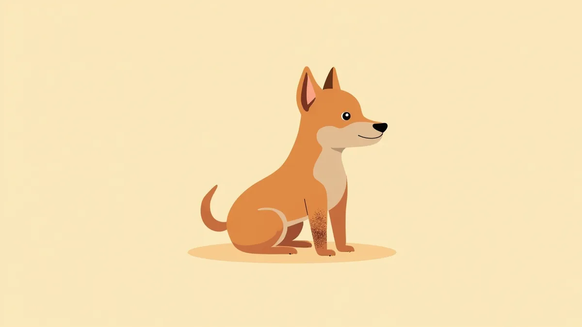
Ringworm symptoms presenting as circular, scaly lesions on a dog's legs
Canine ringworm, or dermatophytosis, is a fungal skin infection caused by dermatophytes, most commonly Microsporum canis. In dogs and cats, the majority of cases are caused by Microsporum canis and Trichophyton mentagrophytes. - Primary Pathogen: Microsporum canis is the most frequently identified dermatophyte in dogs and cats, with cats serving as the primary reservoir hosts. - Transmission: The infection spreads to dogs through contact with spores found on infected animals or in contaminated environments, such as bedding, rugs, and cages. - Symptoms: Common signs include scaling and crusting, often accompanied by circular, hairless lesions. These lesions typically appear on the face, around the mouth, on the ears (pinnae), legs, toes, and tail. The level of itching varies from dog to dog. - Differential Diagnosis: Because ringworm can resemble parasitic infections or other skin conditions, a veterinary evaluation is necessary for an accurate diagnosis. - Prevention: Key preventive measures include cleaning the environment to remove potential sources of infection, caring for your dog’s paws after outdoor activities, and avoiding contact with infected cats.

The main causes are Microsporum canis spores and contaminated environments.

The most common cause of dermatophytosis in dogs is exposure to fungal spores, such as those from Microsporum canis. These spores are frequently transmitted through the fur or skin of infected animals, particularly cats. They can also survive for extended periods in contaminated environments, including bedding, rugs, play areas, and cages. Infection begins when a dog comes into contact with these spores from an infected animal or environment. The condition is more likely to develop in warm, humid conditions, under stressful circumstances, in overcrowded housing, or where hygiene is poor, and it is especially common in puppies. However, without direct exposure, advanced age alone does not increase the risk of infection.
Microscopic fungal spores and hair fragments found on a contaminated dog bed

The main symptoms are circular lesions and hair loss.

The most characteristic sign of canine dermatophytosis is skin lesions accompanied by scales and crusts. These lesions often appear as circular or semicircular patches of hair loss, with the hair in the center falling out to expose the skin. Hair loss typically occurs on the face, around the mouth, ears, legs, toes, and tail. The degree of itching varies from dog to dog; some may experience severe itching, while others may have little to none. Lesions can be localized to a single area or spread throughout the body, and they may appear symmetrically or asymmetrically. As the lesions gradually expand, they can take on a "ringworm" (ring-shaped) appearance.
Progression of ringworm, characterized by multiple circular, scaly lesions on the dog's face and legs.

When to Visit the Vet Immediately

If the lesion spreads rapidly or your dog shows severe itching and repeatedly licks or scratches the area, you should visit the vet immediately. Additionally, since it can spread to other animals or family members, isolation measures are necessary. Puppies and dogs with weakened immune systems are especially at risk of developing complications.

Diagnosis is confirmed through microbiological testing and microscopic examination.
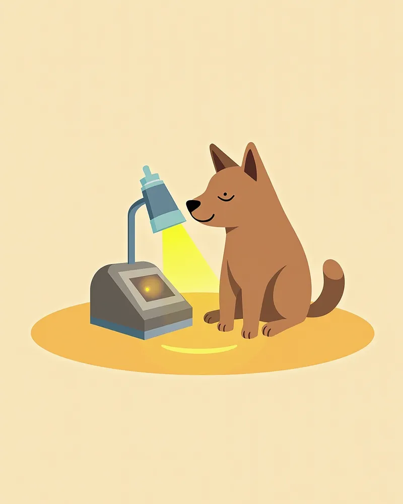
Diagnosing canine dermatophytosis begins with collecting hair and skin scales from the affected area for testing. Common collection methods include brushing the hair with a new toothbrush, plucking hairs, or using the acetate tape impression technique. When illuminated with a Wood’s lamp, hairs infected with Microsporum canis may exhibit an apple-green fluorescence, which is positive in approximately 90–100% of cases. Direct microscopic examination can reveal hyphae or arthroconidia in the hair. However, fungal culture remains the gold standard for definitive diagnosis, and PCR testing may be performed if necessary. For nodular or refractory lesions, a skin biopsy (histopathology) can aid in differential diagnosis, so additional tests may be conducted based on the veterinarian’s assessment.
The veterinarian shines a Wood’s lamp on the puppy’s skin lesions to check for green fluorescence.

Treatment fundamentally involves the concurrent use of topical and oral medications.

Treating canine dermatophytosis typically involves a combination of topical and oral medications. Topical treatments include antifungal shampoos or creams applied directly to the affected areas. For oral therapy, veterinarians prescribe antifungal agents such as ketoconazole (5–10 mg/kg once daily) or itraconazole (5 mg/kg once daily), dosed according to the dog’s body weight; administering these with food improves absorption. Treatment usually lasts at least four weeks and must continue until the infection is completely resolved both clinically and cytologically, even after visible lesions have disappeared. The veterinarian will make ongoing assessments to determine whether the lesions have fully healed.
A veterinarian is treating a puppy with antifungal shampoo.

Home management hinges on environmental cleaning and isolation.

Proper home management of canine ringworm hinges on thorough environmental cleaning and strict isolation of the infected pet. Wash the dog’s bedding, rugs, and play mats in hot water, then disinfect them in a high-heat dryer. Wipe down walls, floors, and window crevices with an antifungal cleaner to eliminate spores. Keep the infected dog separated from other animals during the contagious period, and make sure you wash your hands frequently. Because ringworm is a zoonotic disease that can spread to humans, maintaining strict hygiene is especially important. Regular veterinary check-ups and diligent environmental management are key to preventing reinfection.
While the infected puppy is being treated in an isolated room, the veterinarian is disinfecting the floor.

Precautions to Prevent Recurrence

Even after treatment is complete, if the lesions haven’t fully resolved, there’s a possibility of reinfection. This is especially true for puppies with weakened immune systems or those in stressful environments, where recovery may be slower or relapses more likely. Regular check-ups and proper environmental management are essential. Since accurate diagnosis of dermatophyte infections requires specific testing, consult your veterinarian if needed.

Reviewed by a veterinarian

Dr. Tony — Punnawat Phongkittirak

Dr. Tony — Punnawat Phongkittirak

Veterinarian

A veterinarian who majored in veterinary medicine at Khon Kaen University, Thailand, and completed the IVSA program at North Carolina State University in the United States. Drawing on clinical experience at animal hospitals, he works in the pet healthcare field and is dedicated to building a digital care environment that connects pet parents with veterinarians.

Frequently Asked Questions

Can dog ringworm be transmitted to humans?
Yes, canine dermatophytosis is a zoonotic disease that can be transmitted to humans. Children and individuals with weakened immune systems are particularly susceptible to developing skin lesions. Always wash your hands thoroughly after handling an infected dog, and seek medical attention if you notice any symptoms.
What is the difference between parasitic infections and dermatophytosis?
Parasitic infections and dermatophytosis can present with overlapping symptoms, making it difficult to distinguish between them based on appearance alone. Dermatophytosis commonly manifests as scales, crusts, and circular alopecic lesions. These lesions may appear in a single area or spread throughout the body, and they can be either symmetrical or asymmetrical. The degree of pruritus also varies from dog to dog. Therefore, an accurate diagnosis requires veterinary testing, such as fungal culture.
How long does the treatment period last?
Treatment typically takes at least four weeks. Even after the lesions have disappeared, additional treatment under your veterinarian’s guidance is necessary until complete clinical and cytological healing is achieved to prevent recurrence. Always consult with your veterinarian before deciding to discontinue treatment.
How should I clean the environment at home?
Wash bedding, rugs, and play mats in hot water or disinfect them in a high-temperature dryer. Clean floors and walls with a cleaner containing an antifungal agent, and thoroughly remove dust from hard-to-reach areas like window crevices. It is important to separate and disinfect contaminated items as quickly as possible.
How can I prevent it?
Avoid contact with infected animals, and prioritize paw care after outdoor activities along with regular environmental cleaning. Puppies and immunocompromised dogs are at higher risk of infection, so extra caution is warranted. Regular veterinary check-ups can help detect early signs of the disease promptly.

Share

Related Guides

References

[1] Moriello KA (2019a) Dermatophytosis in cats and dogs: A practical guide to diagnosis and treatment. /n Practice 41, 138-141.

[2] BSAVA Manual of Canine and Feline Dermatology, 4th Ed (2017). Chapter on Dermatophytosis.

[3] Clinical Medicine of the Dog and Cat, 4th Ed (2019). Dermatologic Disorders section.

This information is based on veterinary literature and does not replace diagnosis or treatment. Please consult a veterinarian for specific health concerns.

Metapet Co., Ltd. | CEO: Park Sung-yong | Business Reg. No. 417-88-02562 | Seoul, South Korea | Customer Center

Dog Ringworm vs. Parasites: Symptoms, Diagnosis & Care | Meongsiljang