Like
Share
멍실장
강아지 체리아이(제3안검 탈출증) 증상과 원인, 진단·치료와 관리 포인트 총정리

If a Red Lump Appears in Your Dog’s Eye Corner — Causes, Treatment, and Management Points for Cherry Eye

Eye HealthDiseasesMeongsiljang Veterinary Advisory Board

Cherry eye in dogs is an eye condition where the third eyelid prolapses inward, appearing as a red, fleshy mass. Early diagnosis and appropriate treatment are essential.

Cherry Eye in Dogs Is a Condition Where the Third Eyelid Prolapses Inward
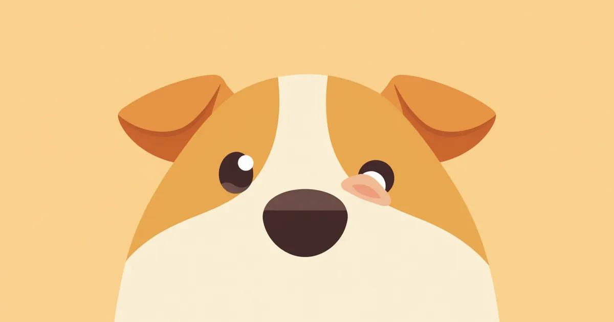
A puppy’s third eyelid has popped out, appearing as a red, fleshy growth.
Cherry eye in dogs is an ophthalmic condition in which the tear gland, located on the inner side of the third eyelid (nictitating membrane), prolapses from its normal position and appears as a smooth, pink-to-red mass protruding in the inner corner of the eye. It primarily affects young puppies and is believed to result from congenital weakness of the connective tissue that supports the third eyelid, allowing the tear gland to slip out of place. The prolapsed gland may have reduced tear production, and it is often accompanied by conjunctivitis or discharge such as eye boogers. Furthermore, exposure to air can dry and irritate the tissue, potentially leading to secondary inflammation, bacterial infection, or corneal ulcers. Because cherry eye is more than just a cosmetic issue and can directly impact eye health, it is important to seek veterinary care for an accurate diagnosis and appropriate management as soon as it is noticed.

The Main Causes of Cherry Eye Are Genetic Predisposition and Ocular Structure
- Genetic predisposition (weakness of connective tissue): Cherry eye is known to occur when the connective tissue supporting the third eyelid is congenitally underdeveloped, causing the gland to loosen and prolapse. It is commonly observed in predisposed breeds such as English Bulldogs, Beagles, Cocker Spaniels, Boston Terriers, Shih Tzus, Lhasa Apsos, Pekingese, and Basset Hounds. - Characteristics of the third eyelid support structure: The third eyelid consists of a T-shaped cartilage and the gland located beneath it. If this supporting structure is weak or the cartilage becomes deformed, the gland is more likely to slip out of place. - Young age: Cherry eye is known to occur primarily in young puppies. For predisposed breeds, it is important to consistently monitor your dog’s eye condition from an early age. - Unilateral or bilateral occurrence: While it may affect only one eye, it can occur in both. Therefore, if it appears in one eye, it is crucial to closely observe and care for the other eye as well.
Diagram of a dog’s third eyelid and supporting tissue structures

The Main Symptoms of Cherry Eye Are a Red Fleshy Mass in the Eye and Frequent Scratching

- Red or pinkish mass: A smooth, pink to red lump may protrude from the inner corner of the eye. This is a prolapsed third eyelid gland, which may initially appear small but can gradually enlarge or persist permanently. - Frequent scratching: Discomfort may cause your pet to frequently scratch or rub the eye, which can worsen irritation and damage to the exposed tissue. - Discharge and conjunctivitis: Ocular discharge, such as that seen with conjunctivitis or crusty eye boogers, is often present. Exposure of the prolapsed gland to air can dry out the surface, increasing irritation. - Redness and reduced tear production: The eye may appear red, and impaired function of the third eyelid gland can lead to decreased tear production. If left untreated for an extended period, this can result in secondary infections or corneal ulcers.
It looks like Cherry is scratching her eyes.

If the Eye Is Red and Swollen with Continuous Tearing, Immediate Veterinary Visit Is Required

After cherry eye occurs, if the eye becomes severely swollen, tears continuously, or the pet cannot open its eye, these are signs of infection or corneal damage. In such cases, you should consult a veterinarian immediately. If left untreated, it can lead to vision loss.

Diagnosis Is Accurately Confirmed by the Veterinarian’s Eye Examination and Ophthalmic Instruments
- Visual Examination (Inspection): Diagnosis is fundamentally based on the veterinarian directly and carefully observing the structure of the third eyelid to confirm whether the tear gland has prolapsed, as well as its location and condition. At this time, the degree of redness, discharge, and swelling is also assessed. - Medical History and Physical Examination: We determine when the symptoms first appeared, whether they affect one or both eyes, and examine the overall condition of the eyes. - Corneal Examination: Fluorescein staining is used to check for secondary corneal ulcers or damage. - Tear Testing: Since a prolapsed tear gland may have reduced tear production, tear function is evaluated through tests such as measuring tear volume if necessary. - Comprehensive Assessment: We synthesize these various findings to determine the severity of the prolapse and the presence of any complications. The earlier the diagnosis, the easier the management and recovery process becomes.
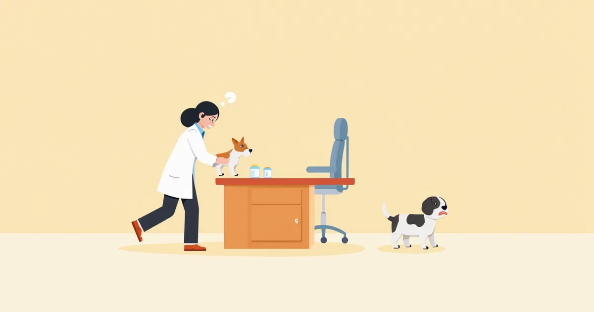
A veterinarian examining a dog's eyes with a slit lamp.

Treatment Is Divided Into Medication Management and Surgery, With Surgery Required in Most Cases

- Medication: In the early stages, anti-inflammatory and antibiotic eye drops can help alleviate inflammation and secondary infections. However, these only provide symptomatic relief and do not address the underlying cause. - Surgery as the primary treatment: The most ideal treatment is surgery to return the prolapsed lacrimal gland to its original position and secure it in place. Excision (removal) of the lacrimal gland should be avoided whenever possible, as it can lead to complications such as dry eye (keratoconjunctivitis sicca). - Surgical techniques: The "Morgan pocket" technique, which involves creating a pocket in the conjunctiva to house and secure the lacrimal gland, is the most widely used method. In difficult cases—such as chronic conditions, severe inflammation, or the need for revision surgery—other techniques may be combined. - Recovery process: A recovery period of 1–2 weeks is required after surgery. During this time, it is important to administer prescribed eye drops, prevent infection, and monitor the condition of the cornea. - Preventing recurrence: Even after surgery, regular check-ups are recommended to monitor for recurrence and assess the health of the opposite eye.
Cherry, a puppy recovering from surgery, is wearing an Erica collar.

Home Care Focuses on Preventing Post-Surgical Infection and Ensuring Stable Recovery

- Elizabethan collar (neck cone) use: Have your dog wear an Elizabethan collar after surgery to prevent self-injury from scratching or rubbing the eyes. This is very important during the recovery process. - Eye drop administration: Use the anti-inflammatory and antibiotic eye drops prescribed by your veterinarian on the scheduled regimen to prevent inflammation and infection. - Periocular care: Gently wipe around the eyes with a soft cloth, but avoid rubbing the surgical site directly. - Monitoring for swelling and bleeding: Contact your clinic immediately if the surgical site becomes severely swollen or starts bleeding. - Activity restriction: Avoid excessive activity during the early recovery phase, and carefully manage walks and other exercises to prevent overexertion. - Regular check-ups: Visit the clinic regularly—for example, 1–2 weeks and 1 month after surgery—to monitor recovery progress and corneal health.
For dogs wearing the Erica collar, keep the area around their eyes clean.

Certain Breeds Have a High Risk of Cherry Eye Recurrence, So Caution Is Needed

English Bulldogs, Beagles, Cocker Spaniels, Boston Terriers, Shih Tzus, Lhasa Apsos, Pekingese, and Basset Hounds are breeds known to be more prone to cherry eye. This condition most commonly affects small to medium-sized dogs, and since it can occur in both eyes, it’s a good idea to have the other eye regularly checked even if one has already been treated.

Reviewed by a veterinarian

Dr. Tony — Punnawat Phongkittirak

Dr. Tony — Punnawat Phongkittirak

Veterinarian

A veterinarian who majored in veterinary medicine at Khon Kaen University, Thailand, and completed the IVSA program at North Carolina State University in the United States. Drawing on clinical experience at animal hospitals, he works in the pet healthcare field and is dedicated to building a digital care environment that connects pet parents with veterinarians.

Frequently Asked Questions

Can cherry eye heal on its own?
In the early stages, the prolapsed tear gland may intermittently pop in and out, but over time it typically remains permanently displaced, making spontaneous resolution unlikely. If left untreated, the exposed tissue can become dry and irritated, potentially leading to conjunctivitis, corneal ulcers, and secondary infections.
How painful is cataract surgery for pets?
Since surgery is performed under anesthesia, your pet won’t feel any pain during the procedure. Afterward, there may be some discomfort, but your veterinarian will prescribe appropriate medications, such as anti-inflammatory and pain-relieving drugs, to manage it.
Is there a chance of recurrence after surgery?
While there is a possibility of recurrence, modern surgical techniques such as the Morgan pocket method, which reposition and secure the tear duct in its proper place, can yield relatively stable outcomes. Post-operative care and regular check-ups are essential.
Should a cherry eye be surgically corrected right away?
If symptoms persist or if irritation or complications develop in the eye, surgery should be considered. Since preserving the tear duct is recommended, it’s best to consult with your veterinarian to determine the appropriate timing.
How much does a cherry eye surgery cost?
Surgical costs vary depending on the breed, clinic, and location. Typically, they range from $70 to $200, and you can adjust the budget in consultation with your veterinarian.

Share

Related Guides

References

[1] Maggs DJ. Third eyelid. In: Maggs DJ, Miller PE, Ofri R, et al. (eds.). Clinical Atlas of Canine and Feline Ophthalmic Disease. 2nd ed. Elsevier; 2020.

[2] Côté E. Clinical veterinary advisor. Dogs and cats. 3rd ed. St. Louis, MO: Elsevier Mosby; 2015.

[3] Ettinger SJ, Feldman EC, Côté E. Textbook of veterinary internal medicine: diseases of the dog and the cat. 8th ed. St. Louis, MO: Elsevier; 2017.

This information is based on veterinary literature and does not replace diagnosis or treatment. Please consult a veterinarian for specific health concerns.

Metapet Co., Ltd. | CEO: Park Sung-yong | Business Reg. No. 417-88-02562 | Seoul, South Korea | Customer Center

Red Lump on Dog’s Eye Corner: Cherry Eye Causes, Treatment & Care Tips | Meongsiljang