Like
Share
멍실장
강아지가 CT 스캔 장비 앞에 누워 있는 모습

Complete Guide to Canine Tumor CT and MRI Staging — When Is Which Test Needed?

Oncology/CancerTop PicksMeongsiljang Veterinary Advisory Board

We’ll walk you through how CT and MRI are used for accurate staging of canine tumors, along with the best management strategies recommended by veterinarians.

What Is Canine Tumor CT and MRI Staging?
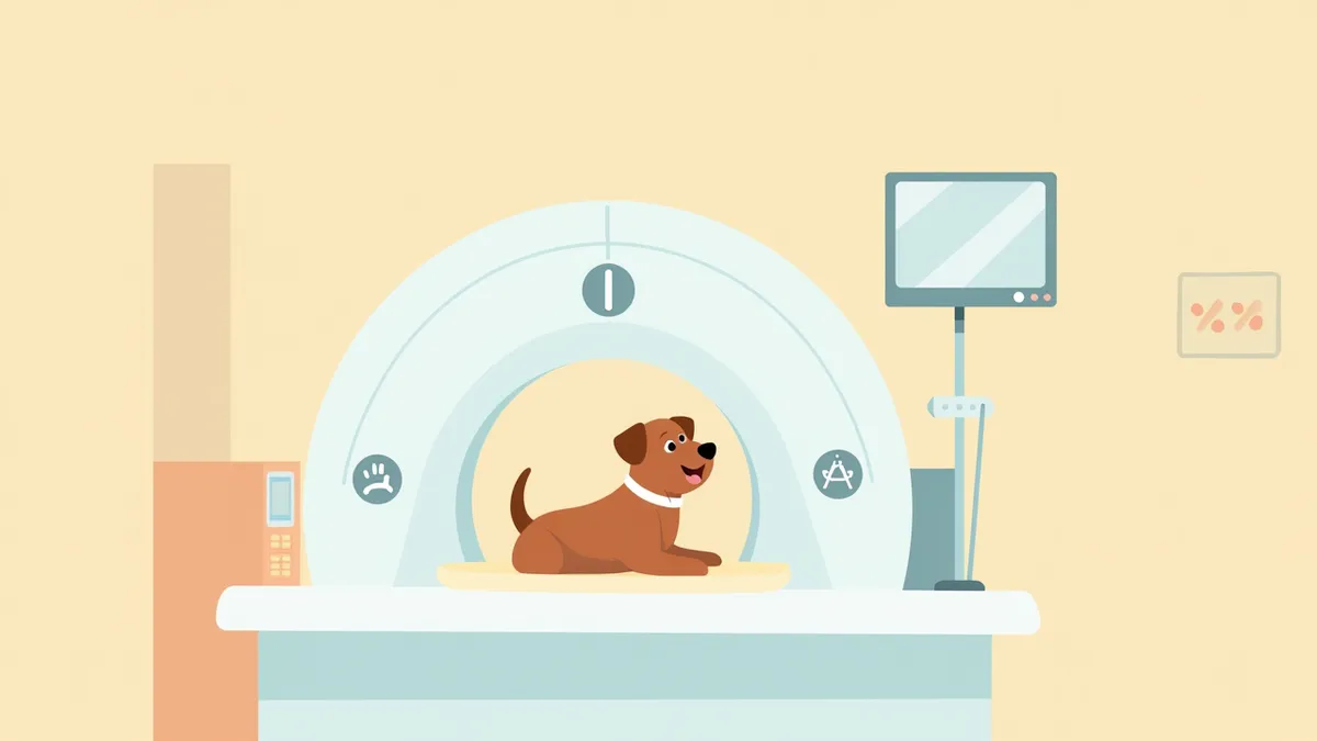
A dog undergoing an MRI scan in a veterinary-supervised imaging suite.
CT and MRI staging are essential imaging tests for precisely determining a dog’s tumor size, location, and whether it has spread to bones or organs. These scans play a crucial role in accurately assessing the cancer stage and guiding treatment decisions. For tumors in the brain, spinal cord, or soft tissues, MRI is preferred because it provides clearer images of soft tissue abnormalities. In particular, MRI is favored over CT for intracranial (brain) lesions, as CT may have lower diagnostic reliability in these cases. On the other hand, CT is advantageous for quickly evaluating bone invasion and lung metastasis, and chest X-rays are often used alongside CT to assess lung involvement. Accurate staging directly influences the planning of surgery, radiation therapy, and chemotherapy. Staging Accuracy: Precisely identifying tumor spread helps determine the most appropriate treatment approach.

Selection Criteria: Tips for Choosing a Hospital for CT and MRI Tests
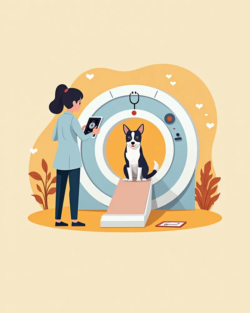
CT scans provide detailed images of bone and lung tissue, while MRI offers more precise visualization of lesions in the brain, spinal cord, and soft tissues. The appropriate imaging modality should be selected based on the tumor’s location.
CT scan: Ideal for assessing bone invasion and detecting lung metastases.
MRI scan: Best suited for evaluating brain and spinal cord tumors, as well as soft tissue involvement.
Combined use: Most effective for a comprehensive assessment of metastatic spread.
A veterinary radiologist analyzing high-resolution MRI images

Check 1: Does the Hospital Offer the Appropriate Test (CT/MRI) for the Tumor Location?
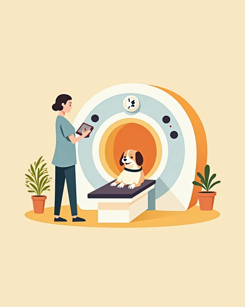
When staging tumors, CT scans are particularly useful for detecting bone involvement and lung metastases, while MRI provides more accurate visualization of brain, spinal cord, and soft tissue lesions. Therefore, it is important to choose the imaging modality that best suits the tumor’s location and suspected metastatic pathways. When selecting a veterinary clinic, verify whether they have the necessary equipment (CT, MRI, or both) and ensure they clearly explain which test is most appropriate for your dog. In some cases, both imaging techniques may be used together to comprehensively assess the presence of metastasis.
A dog undergoing an MRI scan on a 3T MRI machine at a specialized oncology hospital.

Check 2: Is Interpretation Linked with Radiology and Oncology Services?
With CT and MRI scans, accurate interpretation is just as critical as the imaging itself, requiring specialized expertise to correctly analyze the images and link tumor characteristics and staging to an appropriate treatment plan. If a clinic offers integrated oncology services or has dedicated imaging specialists, they can go beyond simply taking the pictures to discuss next steps—such as surgery, chemotherapy, or radiation therapy—based on the results. When choosing a hospital, verify whether imaging interpretation is closely linked with oncology care and whether the staff clearly explains the results so you can fully understand them.
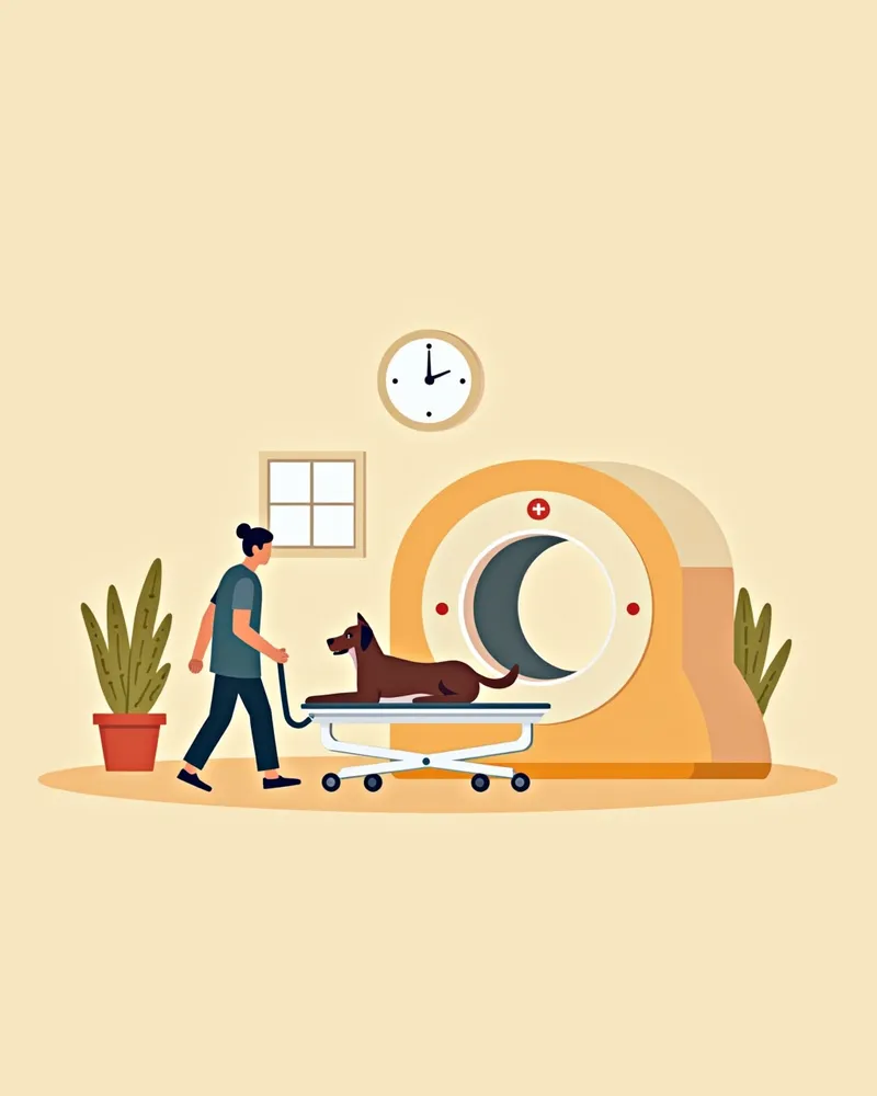
A puppy moving between the hospital’s CT and MRI machines

Check 3: Anesthesia Safety Management, Accessibility, and Consultation Availability

CT and MRI scans require the patient to remain completely still, so anesthesia or sedation is usually necessary. This is especially important for MRI, which takes longer and requires careful management of anesthesia safety. For senior dogs or those with underlying health conditions, a thorough pre-anesthetic evaluation is even more critical, so it’s reassuring to choose a facility with a robust anesthesia monitoring system. If specialized equipment isn’t available locally, you can also opt for a clinic that collaborates with or refers cases to specialized hospitals. Be sure to consider factors like accessibility, scheduling convenience, and how quickly results are communicated.
A dog receiving medical care in a mobile MRI vehicle

Checklist for Choosing a Hospital for Tumor CT and MRI Tests

ItemRecommended criteria
Test suitabilityA facility equipped with CT/MRI suited to the tumor location that guides appropriate testing
Interpretation/coordinationA facility that connects imaging interpretation expertise with oncology care to link through to a treatment plan
Anesthesia safetyA facility with a safety monitoring system for anesthesia/sedation used during testing
Access/collaborative careA facility with a system for referral, collaborative care with specialty hospitals, and result feedback

This is not a table recommending specific products or hospitals, but criteria to reference when consulting your veterinarian. Always make treatment and testing decisions together with your veterinarian through an examination.

Caution: Anesthesia Risks and Pre-Test Preparation

CT and MRI scans usually require anesthesia or sedation to keep your dog still, so it’s important to evaluate their overall health, including heart and lung function, before anesthesia. If your dog is older or has underlying health conditions, a pre-anesthesia evaluation is even more critical. Be sure to discuss this thoroughly with your veterinarian beforehand.

Usage Tips: Pre- and Post-Test Care
Your dog needs to fast for 8 hours before the test. After the procedure, please allow 1–2 hours for recovery from anesthesia. Let the veterinarian know right away if your dog seems different from usual.
Before the test: Fasting, ensuring a calm environment, and completing pre-anesthesia checks.
After the test: Recovery in a quiet space; food can be offered starting 2 hours later.
Records: Be sure to keep the test results, as they will help guide future treatment plans.
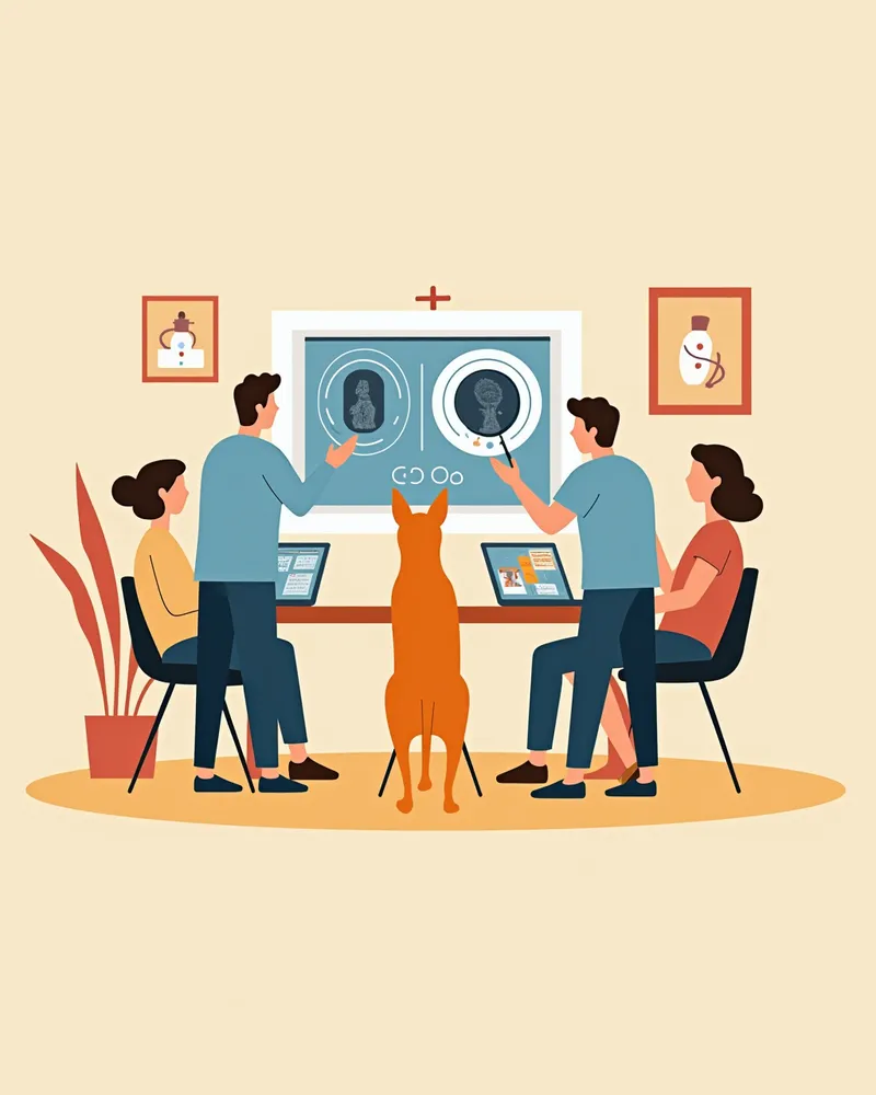
The veterinary oncology team collaboratively reviewing a dog’s imaging scans.

Reviewed by a veterinarian

Dr. Tony — Punnawat Phongkittirak

Dr. Tony — Punnawat Phongkittirak

Veterinarian

A veterinarian who majored in veterinary medicine at Khon Kaen University, Thailand, and completed the IVSA program at North Carolina State University in the United States. Drawing on clinical experience at animal hospitals, he works in the pet healthcare field and is dedicated to building a digital care environment that connects pet parents with veterinarians.

Frequently Asked Questions

When is an MRI necessary for staging tumors in dogs?
MRI excels at soft-tissue contrast, making it highly effective for accurately determining the extent of tumor invasion in the brain, spinal cord, and other soft tissues. However, MRI isn’t necessary for every type of tumor. For lung metastases, chest radiographs (or CT scans) are more appropriate; for bone involvement, CT is preferred; and for intracranial (brain) lesions, MRI is favored due to the lower diagnostic reliability of CT in this area.
Which one is more expensive, CT or MRI?
Costs vary depending on the clinic, the specific tests performed, and your pet’s condition. For an exact quote, please contact the clinic where you plan to have the tests done.
Is it possible to perform CT or MRI scans without anesthesia?
Since movement can blur images and make diagnosis difficult, anesthesia or sedation is usually required. A thorough assessment of the dog’s overall health is essential before anesthesia.
How soon can I receive the test results?
In general, you can expect to receive the results within 1 to 2 days. Specialized clinics may provide results more quickly, while partner clinics may take some time to deliver the results.
How is the treatment plan developed after tumor staging?
Based on the staging results, we comprehensively evaluate the tumor’s size, location, and whether it has metastasized to determine the most appropriate treatment approach—surgery, radiation therapy, or medication.

Share

Related Guides

References

[1] Clinical Medicine of the Dog and Cat, 4th Ed, Chapter 14: Disorders of the Nervous System and Muscle, 2023

[2] Advanced Monitoring for Small Animal Emergency and Critical Care, 2nd Ed, 2021

[3] Veterinary Neuroimaging: MRI and CT in Clinical Practice, 2022

This information is based on veterinary literature and does not replace diagnosis or treatment. Please consult a veterinarian for specific health concerns.

Metapet Co., Ltd. | CEO: Park Sung-yong | Business Reg. No. 417-88-02562 | Seoul, South Korea | Customer Center

Canine Tumor Staging: CT vs MRI — When Is Each Test Needed? | Meongsiljang