
Like
Share
멍실장
강아지 안구 초음파 검사 관리 추천 BEST와 고르는 기준 총정리

Top 7 Recommended Tips for Managing Your Dog Before and After Ocular Ultrasound — Including Selection Criteria

Eye HealthTop PicksMeongsiljang Veterinary Advisory Board

Canine ocular ultrasound is a key diagnostic tool for precisely identifying internal eye abnormalities. To help you manage your pet’s eye health effectively, we’ve summarized the recommended products and selection criteria at a glance.

Canine ocular ultrasound is a key diagnostic tool for precisely identifying internal eye abnormalities.

A veterinarian using an ultrasound probe on a puppy's eye
Ocular ultrasound is a key diagnostic tool that allows non-invasive visualization of the internal structures of your dog’s eye. - High-resolution imaging: Advanced sensors provide clear images of the retina, vitreous body, and intraocular tumors. - Early detection of lesions: It enables early identification of abnormalities not visible externally, ensuring timely treatment. - Minimally stressful procedure: The exam is typically performed while the dog is awake using topical anesthetic eye drops. If your dog is very anxious, sedation may be administered at the veterinarian’s discretion. Since general anesthesia is not required, it is a relatively low-burden procedure. - Accurate diagnostic support: It assists veterinarians in rapidly evaluating various conditions and developing appropriate treatment plans. - Peace of mind for owners: Accurate results allow for systematic management of your dog’s eye health.

6 Key Criteria to Check When Choosing Canine Ocular Ultrasound
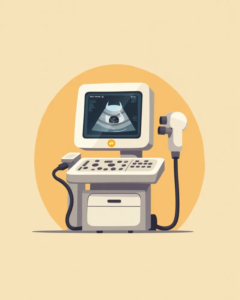
When choosing an ophthalmic ultrasound for your dog, consider the accuracy and reliability of the procedure, as well as convenience for you as the pet owner. Carefully evaluating each factor will help ensure the best possible outcome.
High-precision equipment: Look for devices equipped with high-resolution sensors and advanced image-processing technology.
Experienced professionals: The quality of ultrasound results depends heavily on the technician’s expertise.
A stable examination environment: A quiet, warm space helps keep your dog calm and relaxed.
Prompt result delivery: Immediate diagnostic results help ensure timely treatment.
Reasonable cost: The price should be fair without compromising on quality.
Follow-up care support: Choose a clinic that offers post-exam consultations and ongoing monitoring.
A modern veterinary ultrasound machine displaying a puppy’s eye on the screen.

Check 1: Is high-resolution imaging with an ophthalmic-specific probe available?
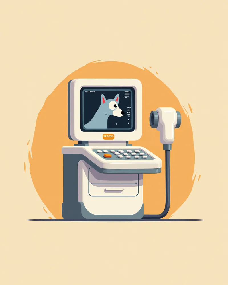
Canine ocular ultrasound is a diagnostic test that visualizes deep intraocular structures such as the retina and vitreous body. To accurately differentiate pathologies like retinal detachment, vitreous hemorrhage, or masses, high-resolution imaging is essential. Low resolution can lead to missed subtle changes or confusion with normal anatomy. When choosing a clinic, verify whether they use a high-frequency probe dedicated to ocular examinations and if the veterinarian has expertise in performing ocular ultrasounds. Reliable results depend on both the quality of the equipment and the examiner’s experience.
An informational image explaining whether high-resolution, eye-specific examinations are available.

Check 2: Can internal structures be visualized when the fundus is not visible?
When cataracts are severe or there is bleeding inside the eye, fundus examination cannot directly visualize the retina, making ultrasound the key tool for assessing internal ocular structures. It is also essential for evaluating retinal health prior to cataract surgery. When choosing a clinic, verify whether they have experience handling such cases and whether they use ultrasound to assess the interior when the fundus is obscured. Clinics with a thorough understanding of appropriate indications can minimize unnecessary tests while providing accurate, essential information.
An illustrative guide showing how to examine the interior when the fundus is not visible

Check 3: Is tracking management possible through measurement and reporting?

To determine the appropriate treatment plan and timing, it is essential to track changes in the size and location of ocular lesions, making it important to keep a record of measurements. Comparing current findings with previous examinations is the only way to know whether the lesion is growing or responding to treatment. When choosing a veterinary clinic, verify whether they accurately measure lesion size and document it in a report, and whether they explain how the current condition compares to previous results during follow-up visits. For more systematic management, consider a clinic that can refer or collaborate with an ophthalmology specialist when needed.
An image illustrating how tracking and management are conducted through measurement and reporting.

Checklist for Selecting a Veterinary Clinic for Ocular Ultrasound

ItemRecommended Criteria
ResolutionA clinic that performs high-resolution eye exams to view retinal and vitreous lesions
Indication ResponseA clinic that examines the interior when the fundus cannot be seen due to cataracts, etc.
Follow-up CareA clinic that manages progression through lesion measurement and reporting
Result InterpretationA clinic where the veterinarian personally explains the exam results and guides the management plan
Cost InformationA clinic that informs you in advance of the estimated cost and the possibility of additional tests before the exam

This is not a table recommending specific clinics, but criteria worth checking when choosing a clinic to perform the exam. Prices and specific programs vary by clinic, so inquire directly before your visit.

Caution: Always assess your dog’s condition before the examination

If your dog is anxious or in pain, the examination may be difficult. Before the exam, the veterinarian will assess your dog’s condition and, if necessary, administer a sedative to ensure the procedure can be performed calmly and safely. In cases of eye trauma or inflammation, treatment must take priority before any examination.

Tips for Canine Ocular Ultrasound

To ensure a successful ocular ultrasound for your dog, keep these key tips in mind: - Rest before the exam: If your dog is stressed or tired, it can be difficult to capture clear images. - Maintain a quiet environment: Prepare a low-noise setting during the procedure to avoid distracting your dog. - The owner’s role is important: It’s best for owners to observe quietly; if your dog becomes anxious, the veterinarian will take steps to help them relax. - Review results immediately: It’s important to examine the images right after the scan and interpret them together with your veterinarian. - Create a follow-up care plan: Based on the results, it’s effective to establish a schedule for ongoing management and monitoring.
A puppy undergoing an ocular ultrasound while in a calm, sedated state.

Reviewed by a veterinarian

Dr. Tony — Punnawat Phongkittirak

Dr. Tony — Punnawat Phongkittirak

Veterinarian

A veterinarian who majored in veterinary medicine at Khon Kaen University, Thailand, and completed the IVSA program at North Carolina State University in the United States. Drawing on clinical experience at animal hospitals, he works in the pet healthcare field and is dedicated to building a digital care environment that connects pet parents with veterinarians.

Frequently Asked Questions

Is an eye ultrasound painful for dogs?
There’s little to no discomfort. Before the exam, we apply topical anesthetic drops, then gently place the probe on the eye’s surface or over the closed eyelid, so your dog experiences minimal discomfort. However, if your dog is very anxious, the veterinarian may take calming measures or use a sedative.
How long does the test take?
The procedure usually doesn’t take long, but the exact time can vary depending on the complexity of the lesion, how cooperative your dog is, and whether sedation is used. It’s best to check with the clinic beforehand for an estimated duration.
Is fasting required before the test?
If the procedure is performed with only topical anesthesia while the dog is awake, fasting is usually not necessary. However, if sedatives might be used because your dog becomes anxious, fasting may be required. Therefore, be sure to confirm with your veterinarian whether fasting is needed before the examination.
What conditions can be diagnosed?
It helps evaluate internal eye lesions that are difficult to examine directly, such as retinal detachment, vitreous hemorrhage or degeneration, and intraocular tumors. It is especially useful for visualizing internal structures when the fundus cannot be seen due to cataracts or hemorrhage.
Are there any special precautions to take after the examination?
After the examination, be careful to avoid irritating the eyes, and follow your veterinarian’s instructions for medication and care.

Share

Related Guides

References

[1] Plumb's Veterinary Drug Handbook, 9th ed. 2022. Wiley-Blackwell.

[2] Textbook of Cardiovascular Medicine in Dogs and Cats, 2021. Elsevier.

[3] Small Animal Ophthalmology, 3rd ed. 2020. Elsevier.

This information is based on veterinary literature and does not replace diagnosis or treatment. Please consult a veterinarian for specific health concerns.

Metapet Co., Ltd. | CEO: Park Sung-yong | Business Reg. No. 417-88-02562 | Seoul, South Korea | Customer Center

Dog Ocular Ultrasound: 7 Pre & Post Care Tips + Criteria | Meongsiljang