Like
Share
멍실장
강아지 치과 방사선 검사 관리 추천 BEST와 고르는 기준 총정리

7 Key Criteria for Dog Dental X-Rays Recommended by Veterinarians

Oral HealthTop PicksMeongsiljang Veterinary Advisory Board

Dental diseases in dogs can include hidden internal issues, so dental X-rays are essential for accurate diagnosis. We’ve compiled a clear overview of our top recommended products and the key criteria for choosing them to help you manage your dog’s dental health effectively.

Dental Radiography is Essential for Diagnosing Dental Diseases in Dogs
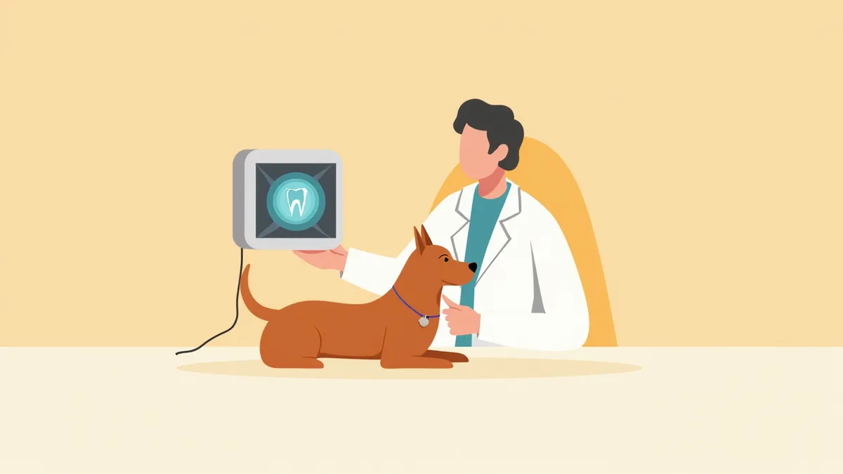
A veterinarian using dental X-ray equipment on a puppy's jaw.
Dental radiography is an essential diagnostic tool that allows for a precise assessment of the condition of your dog’s tooth roots, jawbone, and surrounding tissues. Since evaluation based solely on the visible crowns above the gumline is limited, changes within the roots and alveolar bone can only be detected through radiographic imaging. - Detection of hidden lesions: It enables accurate identification of internal issues not visible to the naked eye, such as root resorption, alveolar bone resorption, retained roots, and periapical abscesses. - Precise imaging under anesthesia: General anesthesia is required to systematically evaluate the entire oral cavity. Digital sensors minimize exposure time, allowing for rapid image acquisition. - Accurate positioning: Positioning aids, such as bite blocks, help maintain consistent positioning to ensure uniform image quality. - Utilization of records and interpretation: Digital images facilitate quick storage and interpretation, aiding in comparisons before and after extractions or treatments. Regular oral examinations enable early detection and preventive care.

5 Recommended Criteria for Dog Dental Radiography
When choosing a dental radiography system for your dog, it’s important to consider factors such as accuracy, safety, and ease of use. By carefully evaluating each aspect, you can select the most suitable imaging method for your pet.
High-resolution images: The system should clearly reveal even subtle changes in the roots and jawbone.
Low-dose radiation: The radiation exposure must remain safe, even with repeated imaging.
Automatic exposure control: Settings should adjust automatically to suit each individual patient.
Mobile equipment: The unit should allow imaging anywhere within the clinic.
Image storage and sharing capabilities: Images should be easily accessible and shareable between the veterinarian and pet owner.
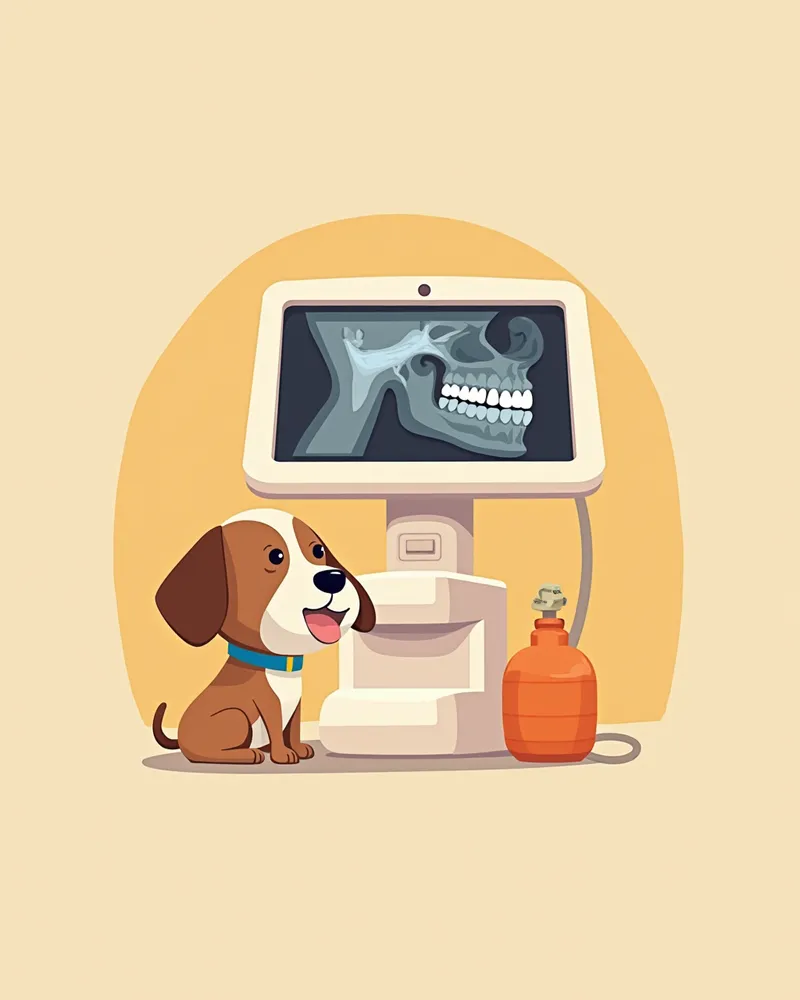
A clear image of a dog's teeth displayed on the monitor of a digital dental X-ray machine

Check 1: Equipped with Digital Low-Dose Radiation Equipment

Dental diseases in dogs often progress beneath the gums, affecting the tooth roots and jawbone, making visual examinations alone insufficient and necessitating dental radiography. Digital sensors allow for quick imaging with lower radiation exposure, minimizing the burden even when multiple areas are captured. When choosing a veterinary clinic, verify whether they are equipped with digital dental radiography systems and whether they perform comprehensive, systematic imaging of the entire oral cavity under general anesthesia. Compared to older film-based equipment, digital systems enable faster re-imaging and interpretation, helping to reduce anesthesia time.
An image providing information on whether we are equipped with digital low-dose radiation equipment.

Check 2: Ability to Detect Subtle Changes in the Jawbone and Tooth Roots

The key to dental radiography lies in detecting subtle changes hidden beneath the gums, such as the tooth roots, alveolar bone resorption, retained root fragments, and periapical abscesses. To visualize these structures clearly, high resolution and precise imaging angles are essential; low-resolution images may miss early lesions, leading to delayed diagnosis until pain has already progressed. When choosing a veterinary clinic, verify whether they use high-resolution imaging to thoroughly evaluate the tooth roots and jawbone, and whether they compare pre- and post-treatment radiographs. This approach helps minimize unnecessary extractions and prevents overlooked treatments.
Guidance images showing whether subtle changes in the jawbone and tooth roots can be detected

Check 3: Image Saving and Sharing for Collaborative Diagnosis

An image explaining whether collaborative diagnosis is possible through saving and sharing medical images
Being able to save and share imaging results is incredibly helpful for comparing progress at follow-up visits or for consulting with other veterinarians on complex cases. Clinics that provide clear diagrams or explanations alongside the images, detailing which teeth have specific issues, make it much easier for pet owners to understand the treatment plan. When choosing a clinic, check whether they maintain and share imaging records with you, and whether they compare new findings with previous records during regular dental cleanings and check-ups. Clinics with thorough record-keeping enable consistent, long-term monitoring of your pet’s oral health.

Checklist for Choosing a Clinic for Dental Radiography

ItemRecommended Criteria
EquipmentA facility equipped with digital low-dose radiography equipment
ResolutionHigh resolution capable of showing fine lesions of the jawbone and tooth roots
Record ManagementA facility where images can be stored and shared to enable progress comparison and collaborative consultation
Result InterpretationA facility where the veterinarian personally explains the test results and guides the management plan
Cost GuidanceA facility that informs you in advance of estimated costs and the possibility of additional tests before the examination

This is not a table recommending specific clinics, but criteria worth checking when choosing a clinic to entrust with the examination. Prices and detailed programs vary by clinic, so inquire directly before visiting.

Caution: Avoid Excessive Repeat Imaging During Radiography

X-ray exams are essential for accurate diagnosis, but unnecessary repeat imaging can lead to excessive radiation exposure. Therefore, X-rays should only be performed when deemed necessary by the veterinarian, and proper radiation protection guidelines must be followed during the procedure to minimize exposure. The frequency and scope of the exams should be determined in consultation with your veterinarian based on your dog's oral health status.

Tips for Dog Dental Radiography
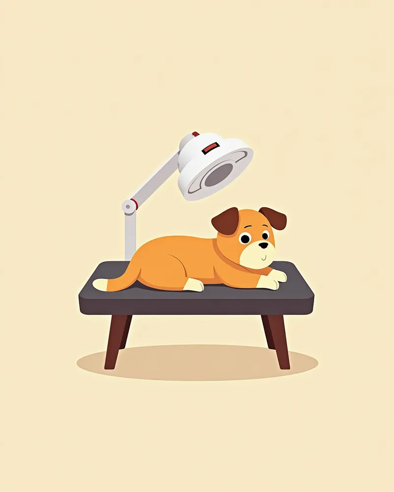
It’s important to help your dog relax in a quiet environment before the exam. - Pre-exam preparation: A short walk or gentle stretching can help release pent-up energy, making your dog more cooperative during the visit. - Comfort during the exam: Dental X-rays are performed under general anesthesia, so having you stay quietly by your dog’s side before anesthesia helps them feel more secure. - Reviewing the images: After the X-rays, carefully review the results with your veterinarian and discuss the treatment plan together. - Regular check-ups recommended: Schedule regular dental exams, and let your veterinarian determine the appropriate frequency of X-rays based on your dog’s dental condition. - Caution with repeated imaging: Avoid excessive X-rays unless specifically recommended by your veterinarian.
A puppy lying quietly during a dental X-ray.

Reviewed by a veterinarian

Dr. Tony — Punnawat Phongkittirak

Dr. Tony — Punnawat Phongkittirak

Veterinarian

A veterinarian who majored in veterinary medicine at Khon Kaen University, Thailand, and completed the IVSA program at North Carolina State University in the United States. Drawing on clinical experience at animal hospitals, he works in the pet healthcare field and is dedicated to building a digital care environment that connects pet parents with veterinarians.

Frequently Asked Questions

Does a dental X-ray cause pain in dogs?
Dental X-rays are performed under general anesthesia, so your pet won’t feel any pain from the procedure itself. Anesthesia is necessary to keep the mouth completely still for accurate imaging of the entire oral cavity, and the veterinarian will closely monitor and manage both the anesthesia and imaging process to ensure your pet’s safety.
How long does the test take?
The duration of the dental X-ray session varies depending on your pet’s oral condition. Digital equipment speeds up both image capture and interpretation, helping to reduce anesthesia time, and the images can be reviewed immediately after they are taken.
Why can’t I get a retest?
Because it’s important to minimize radiation exposure, we only perform X-rays when the veterinarian deems them necessary. While regular dental check-ups are essential, we should avoid unnecessary and excessive repeated imaging without proper justification.
Is it possible to perform X-rays at home?
No. Because it requires specialized equipment, general anesthesia, and a veterinarian’s assessment, this procedure can only be performed at a veterinary clinic.
When will I receive the test results?
After the scan, the veterinarian will analyze the images and show you the results. If needed, they’ll also explain any additional tests or treatment plans.

Share

Related Guides

References

[1] Plumb's Veterinary Drug Handbook, 9th Edition, 2022

[2] Veterinary Dental Radiology: A Practical Guide, 2nd Edition, 2021

[3] American Veterinary Dental College (AVDC) Guidelines on Dental Imaging, 2020

This information is based on veterinary literature and does not replace diagnosis or treatment. Please consult a veterinarian for specific health concerns.

Metapet Co., Ltd. | CEO: Park Sung-yong | Business Reg. No. 417-88-02562 | Seoul, South Korea | Customer Center

Dog Dental X-Rays: 7 Key Vet-Recommended Criteria | Meongsiljang