Like
Share
멍실장
강아지 렌즈 탈구 자주 묻는 질문 Q&A — 보호자가 꼭 알아야 할 핵심

What Is Lens Luxation in Dogs? — Timing for Emergency Surgery to Prevent Blindness

Eye HealthQ&AMeongsiljang Veterinary Advisory Board

Lens luxation in dogs is an emergency condition that can cause sudden vision loss and pain. Here’s the essential information every pet owner should know.

What Is Lens Luxation in Dogs?

A medical image showing a dislocated lens in the front part of a dog's eye
Lens luxation in dogs occurs when the lens shifts from its normal position behind the iris. The lens can dislocate forward into the anterior chamber or backward toward the vitreous body. Partial displacement is referred to as subluxation. This condition can cause vision changes and pain, and may lead to secondary complications such as uveitis, glaucoma, and corneal edema. It is particularly common in terrier breeds with a genetic predisposition, such as Jack Russell Terriers and Sealyham Terriers. Abnormalities in the zonular fibers that hold the lens in place can make displacement more likely. The condition may appear suddenly or progress chronically, making prompt diagnosis and treatment essential for preserving vision.

Why Does Lens Luxation Occur in Dogs?
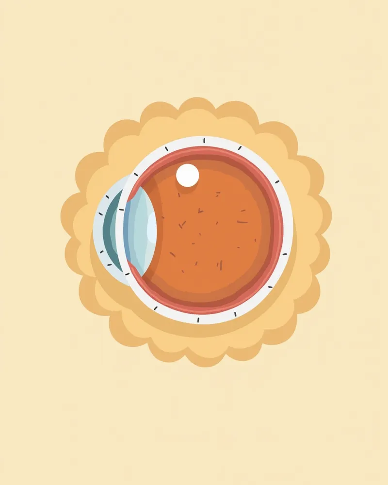
The primary cause of lens luxation is weakening or abnormality of the zonular fibers (suspensory ligaments) that hold the lens in place. Genetic factors, trauma, glaucoma, chronic intraocular inflammation (uveitis), and vitreous degeneration can act alone or in combination to cause this condition. It occurs at a higher rate in terrier breeds with a genetic predisposition, such as Jack Russell Terriers and Sealyham Terriers. When the zonular fibers are compromised, the lens can easily dislocate from its normal position. Lens luxation may appear suddenly as an acute condition or progress gradually as a chronic one, making regular monitoring of your pet’s eye health essential. Early detection significantly improves treatment outcomes.
An anatomical illustration showing the lens and ligaments of a dog's eye

What Are the Main Symptoms of Lens Luxation?

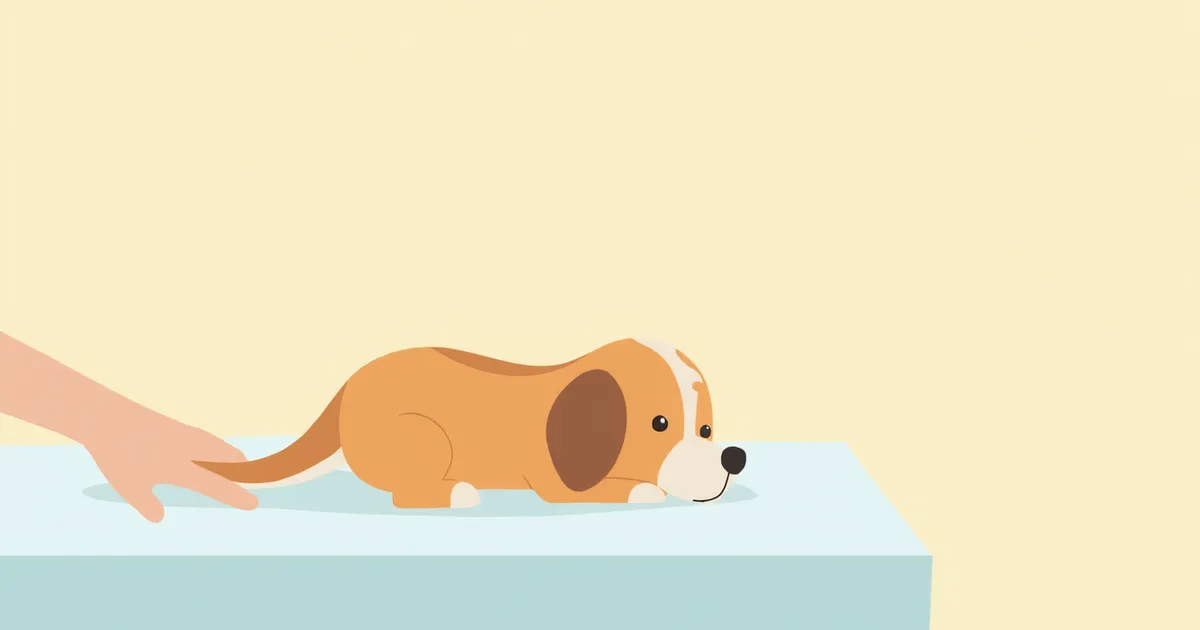
In dogs with lens luxation, sudden changes in vision can occur. When the lens dislocates into the anterior chamber, the cornea may swell and become cloudy (corneal edema), the eye may redden, and pain can develop. Due to pain and light sensitivity, your dog may squint or rub its face. Secondary uveitis or glaucoma can cause further changes in intraocular pressure, worsening symptoms and sometimes making the eyeball appear bulging. Some dogs may bump into obstacles even in familiar environments due to blurred vision. If you notice these signs, seek veterinary care immediately, as prompt treatment is crucial for preserving vision.
A dog experiencing eye pain rubbing its face

Signs That Require Immediate Veterinary Visit

If your pet’s eyes suddenly become red, show light sensitivity, or remain closed, seek veterinary care immediately. Lens dislocation can lead to vision loss, and delayed treatment may make recovery difficult. In particular, severe pain, bulging eyeballs, or persistent tearing are emergency situations.

How Is Lens Luxation Diagnosed?
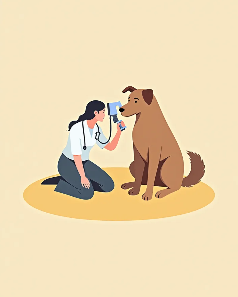
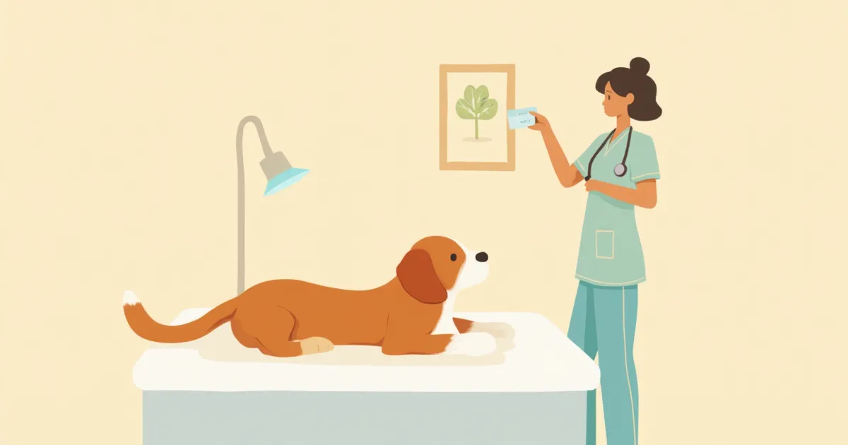
Veterinarians begin with a direct clinical examination of the eye. In cases of severe corneal edema or significant pain, sedation or anesthesia (local or general) may be used to facilitate a thorough evaluation. B-mode ultrasound may also be employed when more precise determination of lens position is needed. Identifying whether the lens has displaced into the anterior chamber or posteriorly is essential for determining the appropriate treatment plan. The veterinarian will also assess for glaucoma and evaluate the severity of inflammation, such as uveitis. A veterinary visit is necessary for diagnosis, and early detection significantly improves treatment outcomes. Professional veterinary assessment is crucial.
A veterinarian examining a dog's eyes using a slit lamp

What Are the Treatment Options?

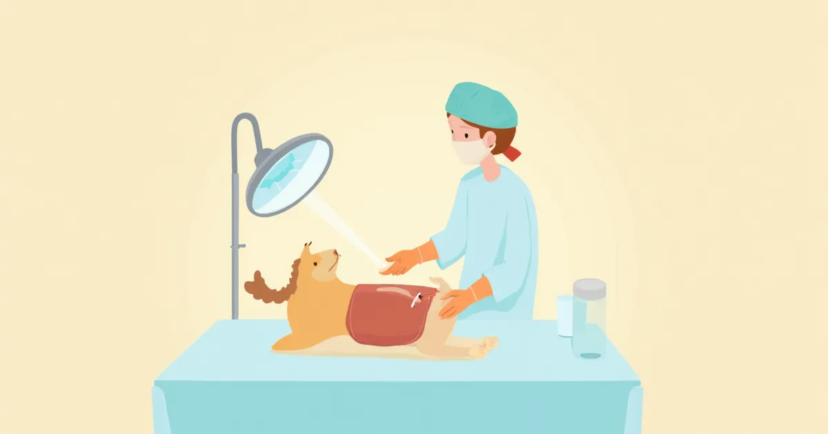
Treatment depends on the position and condition of the lens. In cases of partial dislocation (subluxation), anti-inflammatory medications may be used for management, but surgical removal of the lens is often necessary. Surgery can help restore vision and relieve pain. Long-term anti-inflammatory therapy is typically required after surgery. Additionally, miotics such as demecarium bromide or prostaglandin analogs may be used long-term to prevent the same condition from developing in the other eye. Medications must be used strictly under veterinary guidance, and it is essential to adhere to pre- and post-operative care instructions and regular veterinary check-ups. Early treatment significantly impacts recovery.
A medical illustration showing the surgical process of removing the lens from a dog's eye.

How to Manage at Home?

After surgery, you can use an Elizabethan collar or eye mask to protect your dog’s eyes. Keep your dog’s nails trimmed to prevent scratching, and provide a quiet, undisturbed environment. Administer medications exactly as directed by your veterinarian. Maintain a clean environment to prevent contamination of the surgical site, and avoid wiping the eyes with wet wipes or water. Be sure to keep all scheduled veterinary appointments, and contact your vet immediately if you notice any unusual symptoms. Proper home care plays a significant role in recovery.
A dog wearing a protective eye mask sits quietly.

Comparison of Lens Luxation Treatments

ItemTiming of ApplicationEfficacySide Effects
Medical TreatmentPartial luxation/early stage, for anti-inflammatory purposesHelps control inflammation and painLimited on its own; caution needed when using miotics
Surgical RemovalAfter confirming lens positionCan restore vision and relieve painRisk of surgical complications such as infection
Observation/Conservative ManagementWhen choosing a non-surgical optionEfficacy may be limitedPossibility of progression and complications

In many cases, surgical removal is commonly chosen. Decide after consulting your veterinarian.

Points to Note

Lens luxation can recur. If it has occurred in one eye, the other eye is also at risk, so regular check-ups are essential. Even after surgery, it’s important to monitor your pet’s eye condition closely and visit the vet immediately if any abnormal symptoms appear.

Reviewed by a veterinarian

Dr. Tony — Punnawat Phongkittirak

Dr. Tony — Punnawat Phongkittirak

Veterinarian

A veterinarian who majored in veterinary medicine at Khon Kaen University, Thailand, and completed the IVSA program at North Carolina State University in the United States. Drawing on clinical experience at animal hospitals, he works in the pet healthcare field and is dedicated to building a digital care environment that connects pet parents with veterinarians.

Frequently Asked Questions

Can lens dislocation be completely cured?
Removing the lens through surgery can restore vision. However, in some cases, full recovery may be difficult. Post-operative care is important.
My dog is only having pain in one eye. Could it be a lens dislocation?
If you notice pain, swelling, or vision changes in one eye, it could be a sign of lens dislocation. Immediate veterinary care is necessary.
Can lens luxation be genetic?
Yes, certain breeds have a genetic predisposition. Terriers, particularly Jack Russell Terriers and Sealyham Terriers, are at higher risk. If there is a family history of the condition, extra caution is warranted.
How long does it take after surgery?
Recovery time varies depending on the individual and their condition. After surgery, long-term use of anti-inflammatory medications and regular check-ups are essential, and management should be carried out according to the veterinarian's guidance.
Is lens dislocation very painful?
Yes, lens dislocation can cause pain. It often leads to symptoms such as squinting and rubbing the face.

Share

Related Guides

References

[1] Binder DR, Herring IP, Gerhard T. Outcomes of nonsurgical management and efficacy of demecarium bromide treatment for primary lens instability in dogs: 34 cases (1990–2004). J Am Vet Med Assoc. 2007;231(1):89–93.

[2] Curtis R. Lens luxation in the dog and cat. Vet Clin North Am Small Anim Pract. 1990;20(3):755–773.

[3] Glover TL, Davidson MG, Nasisse MP, Olivero DK. The intracapsular extraction of displaced lenses in dogs: a retrospective study of 57 cases (1984–1990). J Am Anim Pract. 1991;32(2):105–112.

This information is based on veterinary literature and does not replace diagnosis or treatment. Please consult a veterinarian for specific health concerns.

Metapet Co., Ltd. | CEO: Park Sung-yong | Business Reg. No. 417-88-02562 | Seoul, South Korea | Customer Center

What Is Lens Luxation in Dogs? Emergency Surgery Timing | Meongsiljang