Like
Share
멍실장
강아지 안압 측정(토노메트리) 증상과 원인, 진단·치료와 관리 포인트 총정리

Measuring Your Dog’s Intraocular Pressure (Tonometry) — Early Detection of Glaucoma and How to Read the Numbers

Eye HealthDiseasesMeongsiljang Veterinary Advisory Board

Measuring your dog’s intraocular pressure is a key test for the early detection of eye diseases. Abnormal pressure can be an early sign of serious conditions like glaucoma.

What Is Measuring Your Dog’s Intraocular Pressure (Tonometry)?
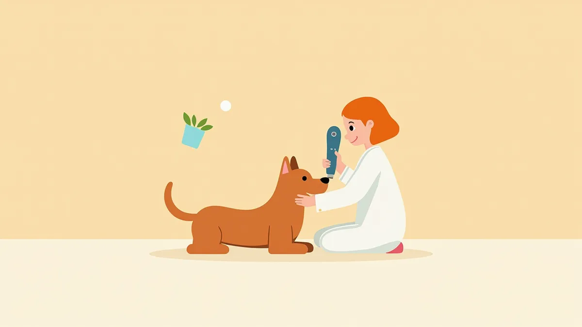
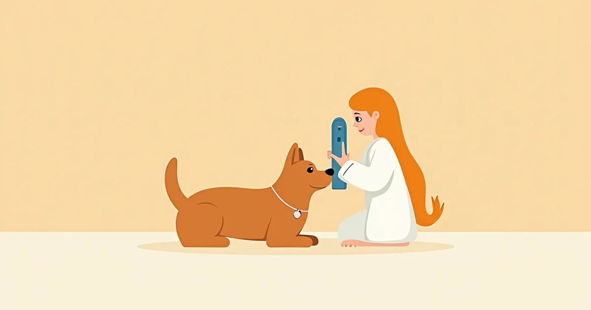
A veterinarian using tonometry equipment on a dog's eye
Measuring your dog’s intraocular pressure is a medical test that accurately assesses the pressure inside the eye, playing a crucial role in the early detection of eye diseases. - Intraocular Pressure Measurement: If the aqueous humor, the clear fluid filling the eye, is not drained properly, pressure can rise and potentially damage the optic nerve. - Tonometry: This test determines intraocular pressure by applying gentle pressure to the cornea or measuring the rebound of a probe. - Normal Range: The normal intraocular pressure for a healthy dog is generally reported to be between 15 and 25 mmHg. - Importance of Early Detection: Abnormal intraocular pressure can be a sign of glaucoma, so regular check-ups are helpful. - The Procedure: The test is completed quickly and involves little to no pain, making it relatively easy for most dogs to tolerate. - When to Test: It is advisable to consult your veterinarian about regular screening if you notice any abnormalities in your dog’s eyes, or if your dog is a breed prone to glaucoma or is senior.

What Are the Main Causes of Elevated Intraocular Pressure?
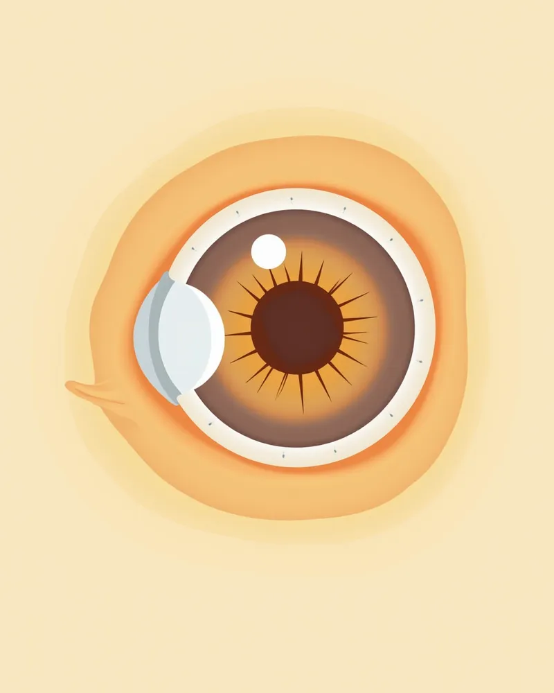
There are various reasons why a dog’s intraocular pressure may increase. - Glaucoma: When the drainage pathway for aqueous humor (the anterior chamber angle) becomes blocked, pressure rises. Primary glaucoma, in particular, is linked to genetic predisposition. - Eye injury: Trauma or impact can disrupt aqueous humor circulation, leading to secondary glaucoma. - Inflammation inside the eye, such as uveitis: Severe or chronic uveitis can cause secondary glaucoma. - Lens luxation, intraocular hemorrhage, or tumors: Physical obstruction of aqueous humor flow can raise intraocular pressure. - Genetic predisposition: Certain breeds, such as American Cocker Spaniels, Basset Hounds, Boston Terriers, Shar-Peis, and Beagles, are at higher risk. - Chronic progression: Gradual degeneration of the anterior chamber angle (trabecular meshwork) over several years can weaken the eye’s ability to regulate pressure.
Diagram of a dog’s eye structure showing increased intraocular pressure due to fluid buildup within the eye

5 Key Symptoms Indicating the Need for Intraocular Pressure Measurement
The following symptoms may indicate the need for intraocular pressure measurement:
Eye swelling: This refers to swelling of the eye or thickening of the eyelids.
Conjunctival redness: This is when the white part of the eye turns red.
Vision loss: This includes behaviors such as stumbling in the dark or bumping into objects.
Excessive tearing: This occurs when tears continuously flow or become thick and sticky.
Squinting or rubbing the eyes: These are signs that the eye may be painful or uncomfortable.
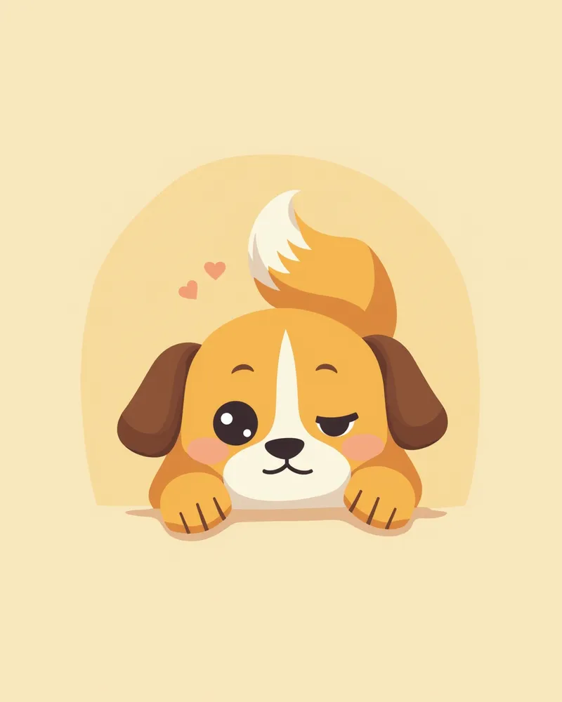
A puppy that seems uncomfortable and is rubbing its eyes

Emergency Signs Requiring Immediate Veterinary Visit

If your pet’s eyes suddenly swell or become cloudy and unresponsive, seek veterinary care immediately. A rapid increase in intraocular pressure can lead to swift vision loss.

Main Diagnostic Methods for Intraocular Pressure Measurement
Intraocular pressure is typically measured using one of two methods: - Applanation tonometry: This method uses a Tono-Pen device to measure pressure by gently touching the cornea. Before the measurement, anesthetic eye drops are applied to the cornea. Multiple readings are taken, and the average value is provided along with the margin of error. - Rebound tonometry: This method uses a Tono-Vet device, where a lightweight probe briefly touches the cornea and analyzes the rebound to determine intraocular pressure. No anesthetic eye drops are required, making it less stressful for the pet. During the test, the pet’s head should be positioned so that the probe is horizontal. Both methods provide quick and safe intraocular pressure measurements. - Criteria for choosing a method: The appropriate method is determined based on the dog’s condition, breed, and overall eye health.
An image comparing the Tono-Pen and Tono-Vet devices

Step-by-Step Treatment Methods for Elevated Intraocular Pressure
When intraocular pressure is elevated, treatment is carried out in stages. - Medication: Eye drops are used to lower pressure by reducing aqueous humor production or enhancing its drainage. Topical carbonic anhydrase inhibitors (dorzolamide, brinzolamide) are commonly used, especially in the early stages. - Surgical intervention: If medication alone fails to control the pressure, surgery to improve aqueous humor outflow may be considered. - Ongoing management: Regular check-ups and consistent medication use are essential. - Treatment goals: The primary aim is to lower intraocular pressure to prevent damage to the retina and optic nerve, preserve remaining vision, and relieve pain. Typically, the goal is to maintain intraocular pressure at or below 19–20 mmHg. - Prognosis: Glaucoma is a progressive disease, so even with management, vision loss or additional interventions may occur. Early detection and consistent care are crucial. - The owner’s role: Adherence to medication and careful monitoring of the eye’s condition are key to successful treatment.
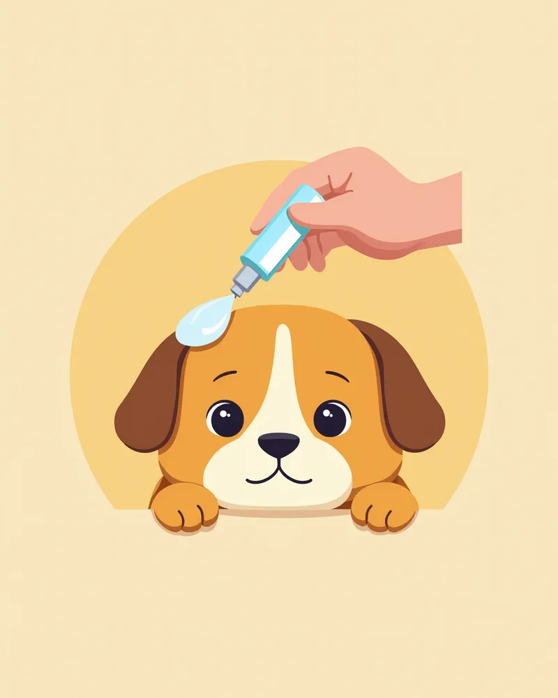
A veterinarian administering eye drops to a puppy

Key Points for Managing Intraocular Pressure at Home
To manage intraocular pressure at home, please follow these guidelines:
Adhere to medication schedules: Administer eye drops exactly at the times prescribed by your veterinarian.
Monitor eye condition: Check daily for swelling, redness, or clouding of the cornea.
Watch for signs of pain or discomfort: Frequent blinking, rubbing, or increased tearing may indicate elevated intraocular pressure and should be taken seriously.
Regular check-ups: Your veterinarian should measure intraocular pressure regularly using a tonometer.
Avoid self-diagnosis: Do not stop or change medications on your own. Consult your veterinarian immediately if you notice any changes.
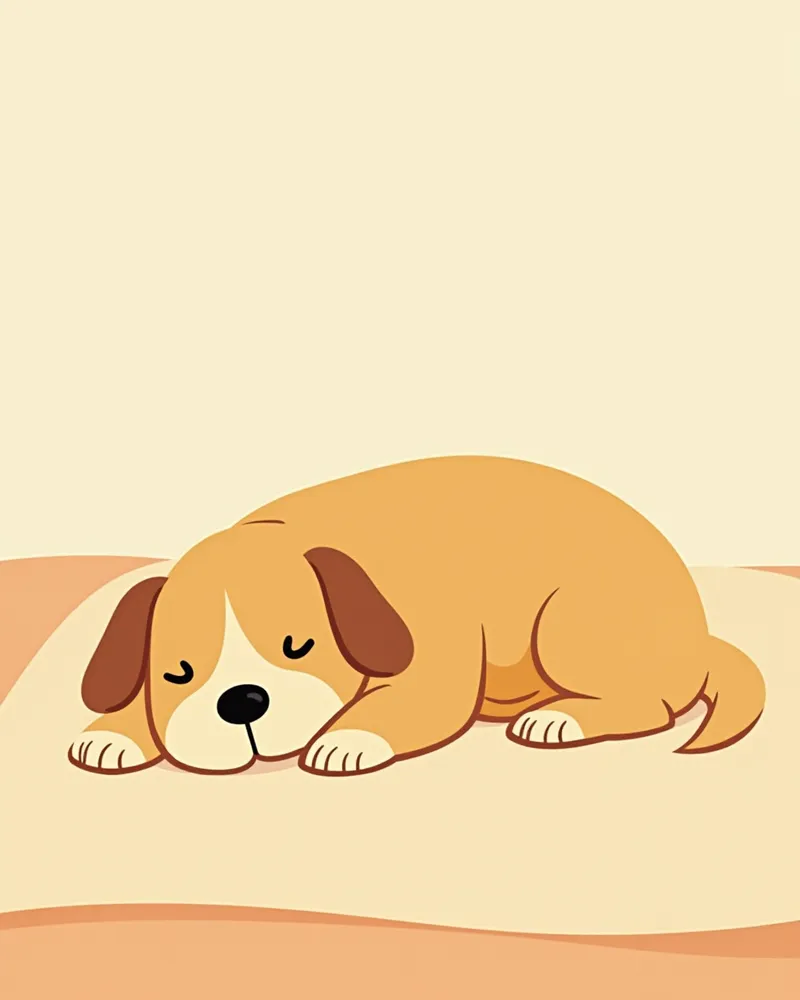
A dog resting comfortably in a calm environment

Certain Breeds Require More Attention to Intraocular Pressure Issues

American Cocker Spaniels, Basset Hounds, Boston Terriers, and Shar-Peis are known to be prone to closed-angle glaucoma, while Beagles and Norwegian Elkhounds are more susceptible to open-angle glaucoma. Additionally, Cairn Terriers, Boxers, and Labrador Retrievers have been reported to have an increased risk of pigmentary glaucoma. Because genetic predisposition plays a role, we recommend regular intraocular pressure checks for dogs of these breeds.

Reviewed by a veterinarian

Dr. Tony — Punnawat Phongkittirak

Dr. Tony — Punnawat Phongkittirak

Veterinarian

A veterinarian who majored in veterinary medicine at Khon Kaen University, Thailand, and completed the IVSA program at North Carolina State University in the United States. Drawing on clinical experience at animal hospitals, he works in the pet healthcare field and is dedicated to building a digital care environment that connects pet parents with veterinarians.

Frequently Asked Questions

Does measuring eye pressure hurt?
In the ablation method, anesthetic eye drops are applied to the cornea beforehand, and since the device only makes light contact, there is minimal discomfort. In the rebound method, a lightweight probe touches the eye only momentarily, so it causes little burden even without anesthesia.
When should intraocular pressure be measured?
If you notice any abnormalities in your dog’s eyes, or if your dog is a breed prone to glaucoma or is senior, it’s best to consult with your veterinarian to determine an appropriate examination schedule. Rather than following a fixed routine, it’s important to tailor the frequency of check-ups to your dog’s individual health status.
Does high intraocular pressure always require surgery?
No. In many cases, intraocular pressure can be managed with medication. Surgery is considered only when medication is no longer effective.
Are there any special precautions to take after measuring intraocular pressure?
After anesthesia, your pet may have difficulty opening their eyes or may rub them. This is usually a temporary occurrence, so there’s no need to worry.
Can dogs with high intraocular pressure play with other dogs?
A little caution is needed. Excessive play can cause trauma to the eyes, so gentle play is recommended.

Comparison of Intraocular Pressure Measurement Methods: Applanation vs. Rebound

ItemApplanation TonometryRebound Tonometry
Device ExampleTono-PenTono-Vet
Topical AnesthesiaRequiredNot required
Measurement PrincipleMeasures the force needed to flatten the corneaMeasures the rebound after a probe momentarily contacts the cornea
Corneal ContactContact until flattening occursOnly momentary contact
Head PositionFew constraintsPositioned so the probe is horizontal

Both methods measure intraocular pressure quickly and safely with handheld devices; applanation requires topical anesthetic eye drops before measurement, while rebound can be performed without anesthesia. The normal intraocular pressure of a healthy dog is reported to be roughly 15–25 mmHg.

Share

Related Guides

References

[1] Gelatt, K.N. & MacKay, E.O. (1998). Distribution of intraocular pressure in dogs. Veterinary Ophthalmology, 1(2–3), 109–114.

[2] Tilley, L.P., Smith, F.W.K. (2007). Blackwell’s Five-Minute Veterinary Consult: Canine and Feline. 4th ed. Ames, IA: Blackwell.

[3] The Veterinary Workbook of Small Animal Clinical Cases. Case 30: The Dog with 'Blue' and 'Red' Eyes. (2020). Elsevier.

This information is based on veterinary literature and does not replace diagnosis or treatment. Please consult a veterinarian for specific health concerns.

Metapet Co., Ltd. | CEO: Park Sung-yong | Business Reg. No. 417-88-02562 | Seoul, South Korea | Customer Center

Dog Tonometry: Detect Glaucoma Early & Read the Numbers | Meongsiljang