Like
Share
멍실장
강아지 눈물길 폐색·비루관 폐색 증상과 원인, 진단·치료와 관리 포인트 총정리

If Your Dog’s Tears Keep Flowing — Causes of Nasolacrimal Duct Obstruction and When to Treat

Eye HealthDiseasesMeongsiljang Veterinary Advisory Board

Nasolacrimal duct obstruction in dogs occurs when tears cannot drain properly, leading to a moist area around the eyes and split hair. The causes can include anatomical abnormalities, inflammation, or trauma.

Canine Nasolacrimal Duct Obstruction Is a Condition Where Tears Cannot Drain Properly

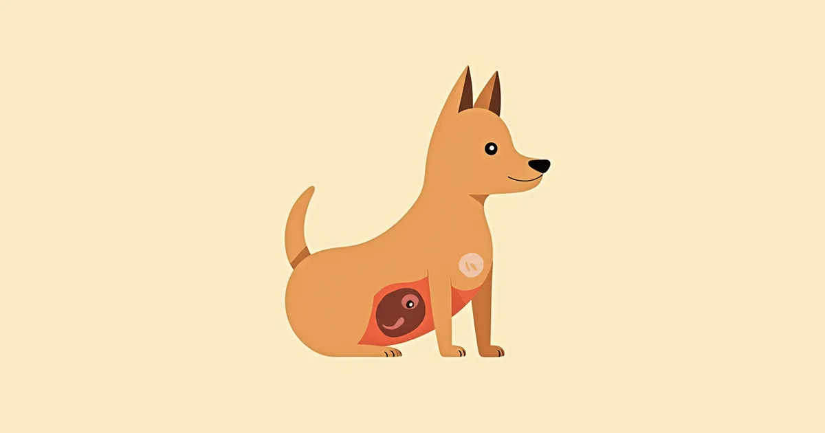
Splitting of the fur around the eyes due to tear duct obstruction
Canine nasolacrimal duct obstruction occurs when tears cannot drain through the nasolacrimal duct into the nasal cavity, causing them to accumulate around the eyes. - Anatomical abnormalities: Brachycephalic breeds with prominent eyes, as well as small breeds like Poodles, often have narrow or kinked nasolacrimal ducts, making tear drainage difficult. - Inflammatory obstruction: Inflammation or bacterial infection around the eyes can block the nasolacrimal duct, leading to tear accumulation. - Traumatic or foreign body obstruction: Tears may fail to drain properly if hair contacts the eye, foreign bodies become lodged, or the eyelid structure is misaligned. If this condition persists, the skin saturated with tears can develop worsening inflammation and hair loss. Early detection and management are essential.

The Main Causes of Nasolacrimal Duct Obstruction Are Anatomical Abnormalities and Inflammation
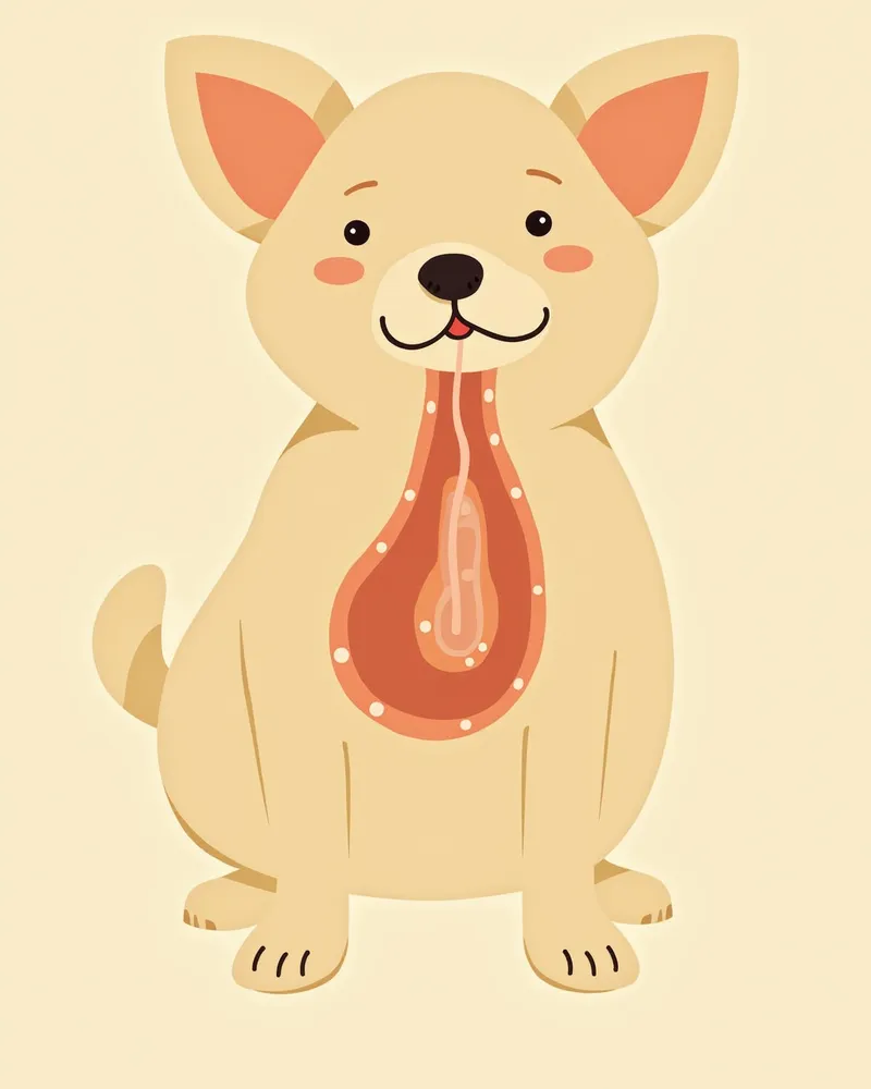
The main causes of tear duct obstruction in dogs are anatomical abnormalities and inflammation of the nasolacrimal duct. - Narrowing of the nasolacrimal duct: In brachycephalic breeds with prominent eyes or small breeds such as Poodles, the nasolacrimal duct may be narrow or kinked. - Inflammatory response: Bacterial infections around the eye or inflammation spreading from adjacent areas can block the nasolacrimal duct, preventing tears from draining properly. - Foreign bodies and trauma: Foreign objects in the eye or malformation of the eyelid structure can obstruct the tear drainage pathway. - Risk of chronicity: If left untreated, post-inflammatory scarring and fibrosis can lead to permanent obstruction, resulting in dermatitis and hair loss. Early diagnosis and treatment of the underlying cause are essential.
Anatomical diagram of a dog's tear ducts

The Main Signs of Nasolacrimal Duct Obstruction Are Persistent Tearing and Split Hair

The hallmark signs of nasolacrimal duct obstruction in dogs include persistent tearing (epiphora) and reddish-brown discoloration of the fur around the eyes. This occurs because tears remain on the skin for prolonged periods, causing irritation and inflammation. Areas where tears accumulate may become stained reddish-brown or develop moist dermatitis, leading to skin maceration. Some dogs repeatedly rub or scratch their eyes, a behavioral response to discomfort. If symptoms persist, complications can affect the eye itself, including the cornea.
Splitting and discoloration of the fur around the eyes due to tear duct obstruction

If Tearing Persists for More Than a Week, Immediate Veterinary Visit Is Necessary

If your pet’s tearing persists for more than a week, or if the skin around the eyes becomes flaky or red, consult your veterinarian immediately. These signs may indicate an infection or chronic inflammation. Left untreated, the eye itself can be damaged, making treatment more difficult. Seek urgent veterinary care if the eyes become red and swollen, or if your pet exhibits intense rubbing or scratching at the eyes.

Diagnosis Is Performed via Tear Flow Test and Endoscopic Examination
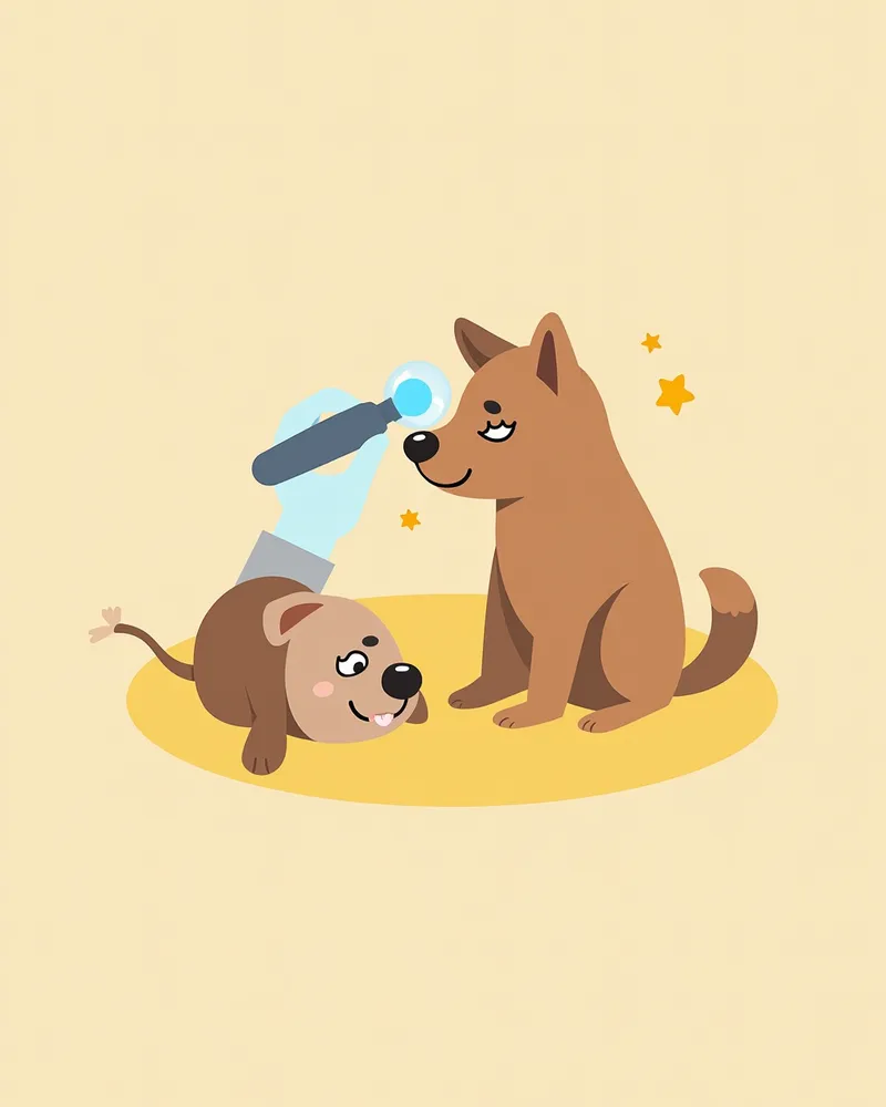
Diagnosing nasolacrimal duct obstruction in dogs begins with the Jones test (fluorescein dye test) to check whether the tear drainage pathway is open. Fluorescein dye is instilled into the conjunctival sac, and if the dye does not appear at the nostril within 10–15 minutes, the nasolacrimal duct is considered obstructed. Bacterial culture and cytology of secretions from the nasolacrimal duct are also performed to confirm the presence of infection. If necessary, contrast radiography or imaging studies such as CT or MRI are used to precisely identify the location of the blockage. Accurate diagnosis is essential for effective treatment.
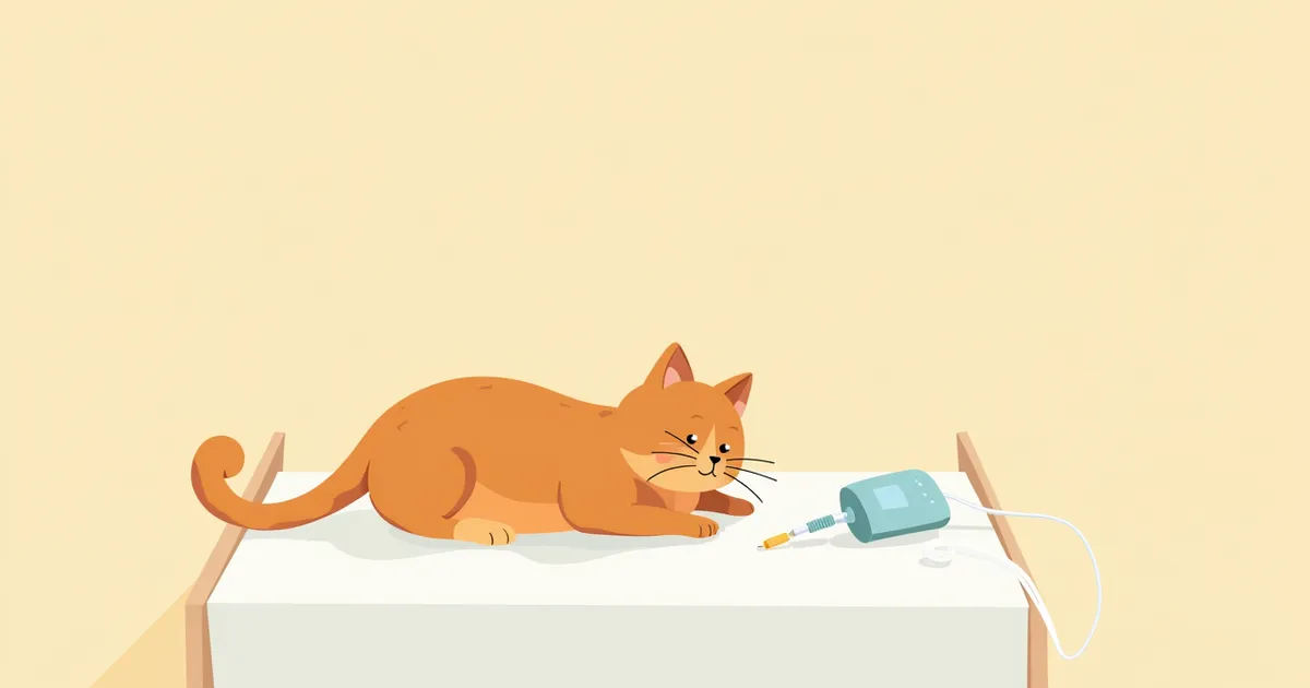
Tear flow test for diagnosing canine nasolacrimal duct obstruction

Treatment Is Carried Out in Stages with Cannulation, Surgery, and Medication

Treatment for canine nasolacrimal duct obstruction is carried out in stages, depending on the underlying cause and severity. - Cannulation, flushing, and stenting: A catheter is inserted into the nasolacrimal duct to clear the blockage and flush the passage, or a stent is placed to maintain patency of the tear drainage pathway. - Medial canthoplasty: This surgical procedure reconstructs the medial eyelid to improve tear drainage and reduce epiphora (excessive tearing). - Medical management: If inflammation or infection is present, topical or systemic antibiotics and anti-inflammatory medications are administered. - Long-term care: Even after procedures or surgery, ongoing cleaning of the periocular area, skin protection, and regular monitoring of corneal health are essential. Effective treatment requires not only addressing immediate symptoms but also implementing preventive care.
Medial canthoplasty during surgery for tear duct obstruction in dogs

Home Care Focuses on Cleaning Around the Eyes and Protecting the Skin

When managing your dog’s nasolacrimal duct obstruction at home, the most important steps are keeping the area around the eyes clean and protecting the skin. Gently wipe around the eyes one to two times a day with a soft cloth. Use only water and avoid any irritating cleansers. Keep areas where tears accumulate dry to help prevent dermatitis. If needed, you can also apply a protective cream recommended by your veterinarian. Proper home care plays a significant role in maintaining the effectiveness of treatment.
Cleaning around the eyes to manage tear duct obstruction in dogs

Breeds with Short Hair and Prominent Eyes, Such as Pekingese and Poodles, Require Attention

Brachycephalic breeds with prominent eyes and small breeds like Poodles are at higher risk for nasolacrimal duct obstruction. Anatomically, these dogs often have narrow or tortuous nasolacrimal ducts, and their puncta may be narrowed or blocked. Therefore, regular cleaning around the eyes and routine veterinary check-ups are essential. If early symptoms appear, prompt intervention is necessary. Prevention is far more important than treatment, so please monitor your pet closely.

Reviewed by a veterinarian

Dr. Tony — Punnawat Phongkittirak

Dr. Tony — Punnawat Phongkittirak

Veterinarian

A veterinarian who majored in veterinary medicine at Khon Kaen University, Thailand, and completed the IVSA program at North Carolina State University in the United States. Drawing on clinical experience at animal hospitals, he works in the pet healthcare field and is dedicated to building a digital care environment that connects pet parents with veterinarians.

Frequently Asked Questions

Is it normal for my pet’s eyes to keep watering?
No, persistent tearing is likely due to a blocked nasolacrimal duct. Since this is a sign of a medical condition, please consult your veterinarian.
Can a blocked tear duct be treated?
Yes, in most cases, treatment is possible through procedures such as catheterization, flushing, stent placement, or medial canthoplasty surgery. The earlier the diagnosis, the easier the management.
Can I treat a blocked tear duct at home?
No, you can’t treat it at home. A veterinarian’s diagnosis is needed to accurately identify the cause of the tear buildup.
Is nasolacrimal duct obstruction hereditary?
Brachycephalic breeds with prominent eyes, as well as small breeds like Poodles, are at higher risk due to their naturally narrow or curved nasolacrimal ducts. However, bacterial infections, inflammation, foreign bodies, and trauma can also cause issues.
Can a blocked tear duct recur after surgery?
While there is a possibility of recurrence, it can be prevented with proper post-operative care and regular check-ups. Please follow your veterinarian's guidance.

Share

Related Guides

References

[1] Gelatt KN et al. (2013) Veterinary Ophthalmology, 2nd ed. Wiley-Blackwell, Oxford, UK.

[2] Esson DW and Calvarese S (2022) Clinical Atlas of Canine and Feline Ophthalmic Disease, 2nd ed. John Wiley & Sons, Inc.

[3] Ny Y et al. (2006) Medial canthoplasty for epiphora in dogs: a retrospective study of 23 cases. J Am Anim Hosp Assoc, 42:435–439.

This information is based on veterinary literature and does not replace diagnosis or treatment. Please consult a veterinarian for specific health concerns.

Metapet Co., Ltd. | CEO: Park Sung-yong | Business Reg. No. 417-88-02562 | Seoul, South Korea | Customer Center

Constant Tearing in Dogs: Nasolacrimal Duct Obstruction | Meongsiljang