Like
Share
멍실장
강아지 렌즈 탈구 증상과 원인, 진단·치료와 관리 포인트 총정리

If Your Dog’s Eyes Suddenly Become Red and Painful — Emergency Signs and Response for Lens Luxation

Eye HealthDiseasesMeongsiljang Veterinary Advisory Board

Canine lens luxation is a condition in which the lens inside the eye shifts out of its normal position, potentially causing pain and vision loss. Early detection and appropriate treatment are essential.

Lens Luxation in Dogs Is a Condition Where the Lens Shifts from Its Normal Position
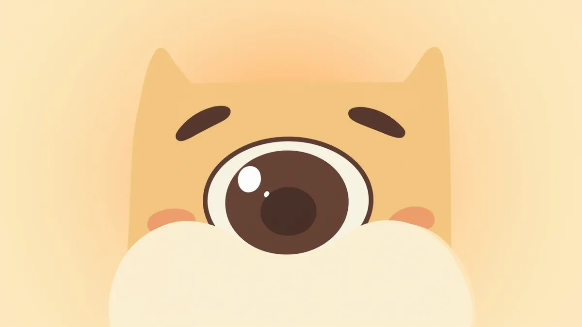
A medical illustration showing a dog’s eye with an anteriorly dislocated lens and corneal edema.
Lens luxation in dogs occurs when the lens shifts from its normal position behind the iris, potentially causing pain and vision loss. This condition can be accompanied by corneal edema, uveitis (inflammation inside the eye), and changes in intraocular pressure (either increased or decreased), making early detection crucial. It is particularly common in several terrier breeds, such as the Sealyham Terrier and Jack Russell Terrier. Primary causes include genetic abnormalities in the zonular fibers that hold the lens in place or in the vitreous body, as well as trauma and chronic intraocular inflammation. When the lens moves into the anterior chamber (the front part of the eye), the cornea may become cloudy, and the dog may rub or squint its eyes due to pain. Severe vision loss can lead to unsteady gait in dim lighting or an inability to see objects. Early diagnosis and appropriate treatment are key to preserving vision.

The Main Causes of Lens Luxation Are Genetics, Trauma, and Increased Intraocular Pressure
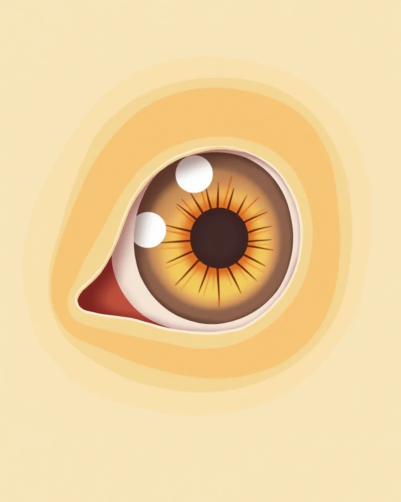
The primary cause of lens luxation is genetic predisposition, with a higher incidence in terrier breeds such as the Sealyham Terrier and Jack Russell Terrier. This genetic factor manifests as structural abnormalities in the zonular fibers that hold the lens in place or in the vitreous body. When these supporting structures weaken, it can lead to luxation. Eye injuries from trauma or impact can also cause lens luxation. Additionally, chronic intraocular inflammation (uveitis), glaucoma, or vitreous degeneration (liquefaction) can weaken the structures that stabilize the lens, resulting in luxation. Because these various factors increase the risk, regular ophthalmic examinations are essential for early prevention.
A medical illustration showing the weakened structure of a dog's eye where the lens-supporting fibers have become fragile.

Main Symptoms Include Tearing, Redness, Pain, and Vision Loss

The main symptoms of lens luxation in dogs include excessive tearing and redness of the eye. If your dog repeatedly rubs or squints their eyes due to pain, it requires attention. A decline in vision may cause unsteady walking in dim light or an inability to see objects. In severe cases, a sudden spike in intraocular pressure can cause the eye to swell or the cornea to become cloudy. This can lead to vision loss, so an immediate veterinary visit is essential.
A photo showing a dog’s red, swollen eye and cloudy cornea due to lens luxation.

Emergency Signs Requiring Immediate Veterinary Visit

If your pet’s eyes suddenly swell, tear up excessively, or if they repeatedly squint or rub their eyes, seek veterinary care immediately. These signs may indicate that the lens has shifted forward, leading to increased intraocular pressure and corneal edema. Anterior lens luxation is a painful, vision-threatening emergency, so if you notice these symptoms, do not delay—get an ophthalmic evaluation as soon as possible.

Diagnosis Is Accurately Confirmed Through Ophthalmic Examination and Ultrasound
Diagnosing lens luxation primarily involves a veterinary ophthalmic clinical examination. If corneal edema is severe or pain makes the exam difficult, veterinarians may use sedation, anesthesia, or B-mode ultrasound to help determine the lens position. In anterior luxation, the lens may be visible in the front chamber of the eye (the anterior chamber), while in posterior luxation, the lens shifts out of the visual axis, often revealing a characteristic finding called an aphakic crescent. Ultrasound can also be used when needed to more clearly assess the lens’s location. For an accurate diagnosis, it is essential to have a professional ophthalmic examination at the clinic.
Ultrasound image of a dog's eye showing a lens dislocated into the anterior chamber

Treatment Is Divided into Surgery and Medication, Selected Based on the Situation

Treatment for lens luxation is divided into surgical removal and medical management. Surgical removal is more common for anterior luxation, accompanied by anti-inflammatory therapy to control inflammation. Posterior luxation or partial luxation (subluxation) may be managed medically with anti-inflammatory drugs (and miotics if needed) while monitoring the condition, or surgically removed if necessary. Since luxation in one eye can lead to the same problem in the other eye, miotics such as demecarium bromide or latanoprost are sometimes used long-term in the unaffected eye for preventive purposes. Treatment approaches vary depending on individual circumstances, so consulting with a veterinary specialist is essential. Early treatment plays a significant role in preserving vision.
A medical illustration showing the surgical process of removing the lens from a dog's eye

Home Care Focuses on Pain Relief and Maintaining Rest

After treatment, it’s important to prevent your puppy from rubbing its eyes at home. Keep the eyes closed and allow for rest, and ensure medications are administered exactly as prescribed. Maintain a quiet, dim environment to reduce stress, and avoid loud noises or bright lights that could irritate the eyes. Restricting your puppy’s movement is also crucial to prevent injury or recurrence. Additionally, regular veterinary check-ups are essential to monitor treatment progress and promptly identify any changes in condition. Pain relief and comfort should be your top priorities.
A dog wearing an eye mask resting comfortably in a dark room

Certain Breeds Are at Higher Risk for Lens Luxation and Require Attention

Terrier breeds, including the Sealyham Terrier and Jack Russell Terrier, have a genetic predisposition that puts them at high risk for primary lens luxation. Dogs of these breeds or those showing any signs of eye abnormalities should undergo regular ophthalmic examinations. If you notice your dog rubbing its eyes, or if you observe redness or pain, seek veterinary care immediately.

Reviewed by a veterinarian

Dr. Tony — Punnawat Phongkittirak

Dr. Tony — Punnawat Phongkittirak

Veterinarian

A veterinarian who majored in veterinary medicine at Khon Kaen University, Thailand, and completed the IVSA program at North Carolina State University in the United States. Drawing on clinical experience at animal hospitals, he works in the pet healthcare field and is dedicated to building a digital care environment that connects pet parents with veterinarians.

Frequently Asked Questions

Can a dislocated lens be completely cured?
In cases of anterior lens luxation, surgically removing the lens can help reduce pain and stabilize the eye. However, depending on the extent of damage that has already occurred, vision may be difficult to restore, so outcomes vary based on individual circumstances.
Can lens dislocation recur?
If one eye experiences a dislocation, the other eye may develop a similar issue. Therefore, preventive medication may be used on the unaffected eye, and regular check-ups and care are essential.
Can it be treated with medication alone?
Medications (such as anti-inflammatory drugs and miotics) help control inflammation and intraocular pressure. While surgical removal of the lens is more common for anterior lens luxation, posterior or partial luxations can sometimes be managed with medication. Your veterinarian will determine the appropriate treatment based on the type and severity of the luxation.
How long does the recovery period after surgery take?
Recovery time varies depending on each pet’s individual condition and the extent of the surgery. After the procedure, it’s important to administer prescribed medications exactly as directed and keep up with regular check-ups on the schedule set by your veterinarian to monitor progress.
Is lens dislocation painful?
The pet is in severe pain, exhibiting behaviors such as rubbing or squinting the eyes, loss of appetite, and reduced activity. Since the pain is acute, immediate action is necessary.

Comparison of Lens Luxation Treatment Methods

ItemTreatment MethodTiming of ApplicationEfficacySide Effects
Surgical Lens RemovalAnterior luxation, elevated intraocular pressureHighPain relief, vision stabilizationPossible infection, hemorrhage, re-luxation
Medical Treatment (Intraocular Pressure Control)Posterior luxation, when surgery is not possibleModeratePain relief, intraocular pressure controlPossible side effects with long-term use
Observation and Follow-up ManagementStable condition, when asymptomaticLowPrevention of recurrenceRequires continuous monitoring

The treatment method is determined by the veterinarian according to the individual situation. Professional veterinary care must be sought.

Share

Related Guides

References

[1] Binder DR, Herring IP, Gerhard T. Outcomes of nonsurgical management and efficacy of demecarium bromide treatment for primary lens instability in dogs: 34 cases (1990–2004). J Am Vet Med Assoc. 2007;231(1):89–93.

[2] Curtis R. Lens luxation in the dog and cat. Vet Clin North Am Small Anim Pract. 1990;20(3):755–773.

[3] Glover TL, Davidson MG, Nasisse MP, Olivero DK. The intracapsular extraction of displaced lenses in dogs: a retrospective study of 57 cases (1984–1990). J Am Anim Pract. 1991;32(2):123–130.

This information is based on veterinary literature and does not replace diagnosis or treatment. Please consult a veterinarian for specific health concerns.

Metapet Co., Ltd. | CEO: Park Sung-yong | Business Reg. No. 417-88-02562 | Seoul, South Korea | Customer Center

Sudden Red, Painful Dog Eyes: Lens Luxation Emergency Signs | Meongsiljang