
If Your Dog’s Eyelids Are Drooping — Causes of Ectropion and Surgical Timing
Canine ectropion is a condition in which the eyelid turns outward, exposing the eye and increasing the risk of pain and infection, making early diagnosis important. It is primarily associated with breed-specific traits and genetic factors.
Canine Ectropion Is a Condition in Which the Eyelid Turns Outward, Exposing the Eye
The Main Causes of Ectropion Are Genetic Factors and Breed Characteristics

Do Not Miss Key Symptom Signals
Immediate Veterinary Visit Is Necessary If Inflammation or Ulcers Develop in the Eye
If your pet’s eyes become suddenly red, painful, excessively watery, or if they keep their eyes closed, it may indicate an infection or ulcer. Because this condition can impair vision, you should consult a veterinarian immediately. Delaying treatment can lead to serious complications.
Diagnosis Is Performed Through the Veterinarian’s Eye Examination and Specialized Instruments
Treatment Is Divided Into Medication and Surgery, Selected Based on the Condition
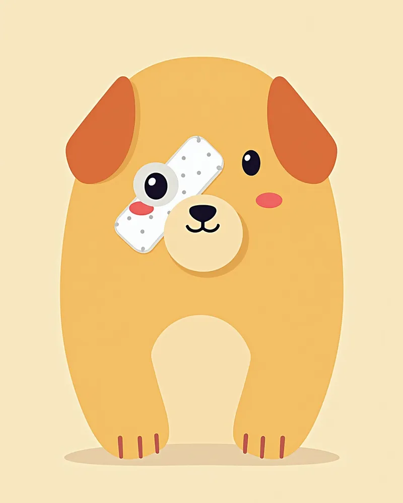
Home Care Focuses on Eye Hygiene and Protection
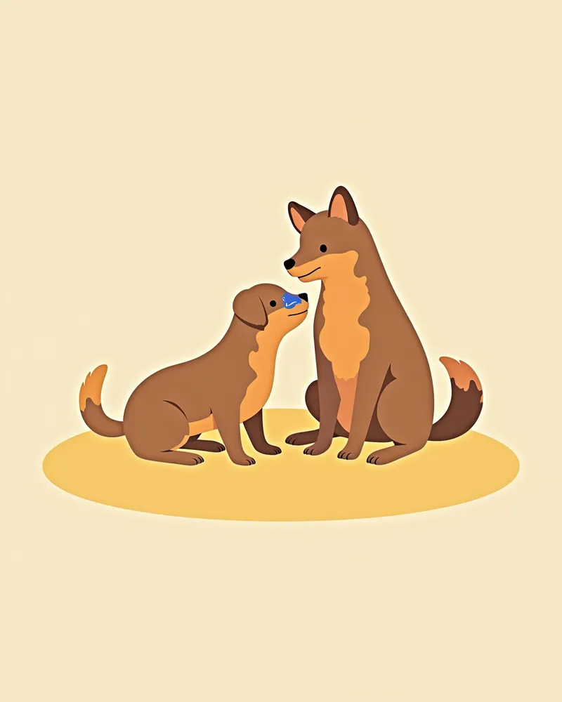
Certain Breeds Have a Genetic Predisposition, So Pre-Adoption Screening Is Important
Breeds with loose facial skin and floppy eyelids, such as Bloodhounds, Saint Bernards, Newfoundlands, and Cocker Spaniels, are at a relatively higher risk of developing ectropion. Since ectropion can have a genetic (congenital) predisposition, it is advisable to review the health records of the parent dogs before adoption and consult with a veterinarian about any potential hereditary conditions. Early detection and consistent care are key to maintaining your pet’s eye health.
Reviewed by a veterinarian

Dr. Tony — Punnawat Phongkittirak
VeterinarianA veterinarian who majored in veterinary medicine at Khon Kaen University, Thailand, and completed the IVSA program at North Carolina State University in the United States. Drawing on clinical experience at animal hospitals, he works in the pet healthcare field and is dedicated to building a digital care environment that connects pet parents with veterinarians.
Reviewed 2026-06-03
Frequently Asked Questions
Is ectropion untreatable?
How long does the surgery take?
Can it come back after surgery?
Can it be managed with medication alone?
Is ectropion painful?
Comparison of Ectropion Treatment Methods
| Item | Treatment Method | Efficacy | Cost | Recovery Period |
|---|---|---|---|---|
| Eye Drops/Artificial Tears | Temporary protection | Low | Inexpensive | Immediate |
| Surgical Treatment | Correction for secondary lesions | High | Moderate to High | 1–2 weeks |
| E-collar (Elizabethan collar) | Prevents self-injury after surgery | Moderate | Moderate | Ongoing |
Ectropion is managed with lubricating and protective eye drops when mild or without complications, and surgical correction is performed when there are significant secondary lesions such as keratitis or corneal ulcers caused by exposure. Surgery is best done after the facial structures have matured, and eye drop therapy is used for symptom relief and corneal protection.
Share
Related Guides

Top 5 Dog Tear Supplements Recommended for 2026

What is Lutein? Effects on Dog Eye Health

The Best Canine Uveitis Management Products and Selection Criteria: A Comprehensive Guide
Dog Foreign Body Obstruction: Symptoms, Causes, Diagnosis, Treatment, and Care Essentials

Canine Megaesophagus: Symptoms, Causes, Diagnosis, Treatment, and Management Essentials
Hydrocephalus in Dogs: Symptoms, Causes, Diagnosis, Treatment, and Management Essentials
If Your Cat Tilts Its Head and Stumbles — Causes, Diagnosis, and Emergency Response for Vestibular Syndrome
Everything You Need to Know About Feline Epilepsy: Symptoms, Causes, Diagnosis, Treatment, and Care
Canine Epileptic Seizures: Symptoms, Causes, Diagnosis, Treatment, and Key Management Points

Feline Corneal Ulcers — Emergency Care and Treatment Options Guide
References
[1] Gelatt KN, Gelatt JP. (1995). Handbook of Small Animal Ophthalmic Surgery: Extraocular Procedures. Pergamon Press.
[2] Curtis R. (1990). Lens luxation in the dog and cat. Veterinary Clinics of North America: Small Animal Practice, 20(3), 755–773.
[3] Ny Y et al. (2006). Medial canthoplasty for epiphora in dogs: a retrospective study of 23 cases. Journal of the American Animal Hospital Association, 42(5), 435–439.