Like
Share
멍실장
강아지 편평세포암 종양학 자주 묻는 질문 Q&A — 보호자가 꼭 알아야 할 핵심

What is Canine Squamous Cell Carcinoma? — Early Signs and Responses You Should Not Miss

Oncology/CancerQ&AMeongsiljang Veterinary Advisory Board

Canine squamous cell carcinoma is a malignant tumor that arises in the skin and mucous membranes, with early detection and appropriate treatment significantly impacting survival rates. We’ve compiled the key questions and answers every pet owner should know.

What is the Tumor Biology of Canine Squamous Cell Carcinoma?

Image showing cellular invasion in a dog's squamous cell carcinoma of the skin
Canine squamous cell carcinoma is a malignant tumor that arises from the keratinocytes of the skin, commonly developing in areas with sparse hair that are frequently exposed to sunlight. In dogs, it most often occurs on the trunk, scrotum, and hind legs, while it is relatively rare around the lips, nose, and anus. - Abnormal changes in sun-exposed skin areas: Breeds with short, white, or spotted coats, as well as those with lightly pigmented skin, are at higher risk due to sun exposure. - The importance of early detection: Small, localized lesions have a favorable prognosis when treated early, but because metastasis can occur, early diagnosis is critical for survival. - Characteristics of the lesions: Shallow, crusted ulcers that deepen into crater-like shapes, or nodular growths accompanied by erosion and bleeding, require immediate veterinary examination. - Treatment success rate: When lesions are small and localized, complete surgical excision can lead to a good prognosis. Early diagnosis and appropriate treatment are key.

What Are the Main Causes of This Disease?

The primary cause of squamous cell carcinoma in dogs is prolonged exposure to ultraviolet (UV) radiation. Dogs with short coats and lightly pigmented skin (white or piebald) are especially vulnerable to UV damage. - UV Exposure: Repeated or long-term UV exposure to unpigmented skin increases risk, so it’s best to avoid extended outdoor activities during peak sunlight hours. - Breed and Skin Color: Sun-induced squamous cell carcinoma is more common in breeds with short coats and white or piebald skin, such as English Bull Terriers, Dalmatians, and Beagles (no specific gender predisposition has been identified). - Chronic Irritation: Recurrent skin injuries or chronic changes such as ulcers and scabs may also be associated with the condition. - Papillomavirus: Papillomavirus involvement has been identified in some cases of squamous cell carcinoma. To help prevent this condition, consider using sun protection or protective clothing.
A puppy exposed to sunlight

What Symptoms Appear?

Canine squamous cell carcinoma often begins with crusty, scaly changes in the skin. Over time, it can grow into nodules or develop into ulcers. In dogs, it most commonly appears on the trunk, scrotum, and hind legs, and while it’s less common on the lips and nose, it can occur anywhere on the body that’s frequently exposed to sunlight. If symptoms persist or worsen, a prompt veterinary visit is essential.
Skin ulcers: Shallow, crusted ulcers that fail to heal and deepen into crater-like lesions.
Thickened skin or nodules: Areas that become thickened and raised, resembling nodules or warts.
Erosion and bleeding: Redness, erosion, crusting, and possible bleeding at the affected site.
Pain response: Signs of discomfort or pain when the area is touched.
Increase in size: If the lesion continues to grow, veterinary evaluation is necessary.
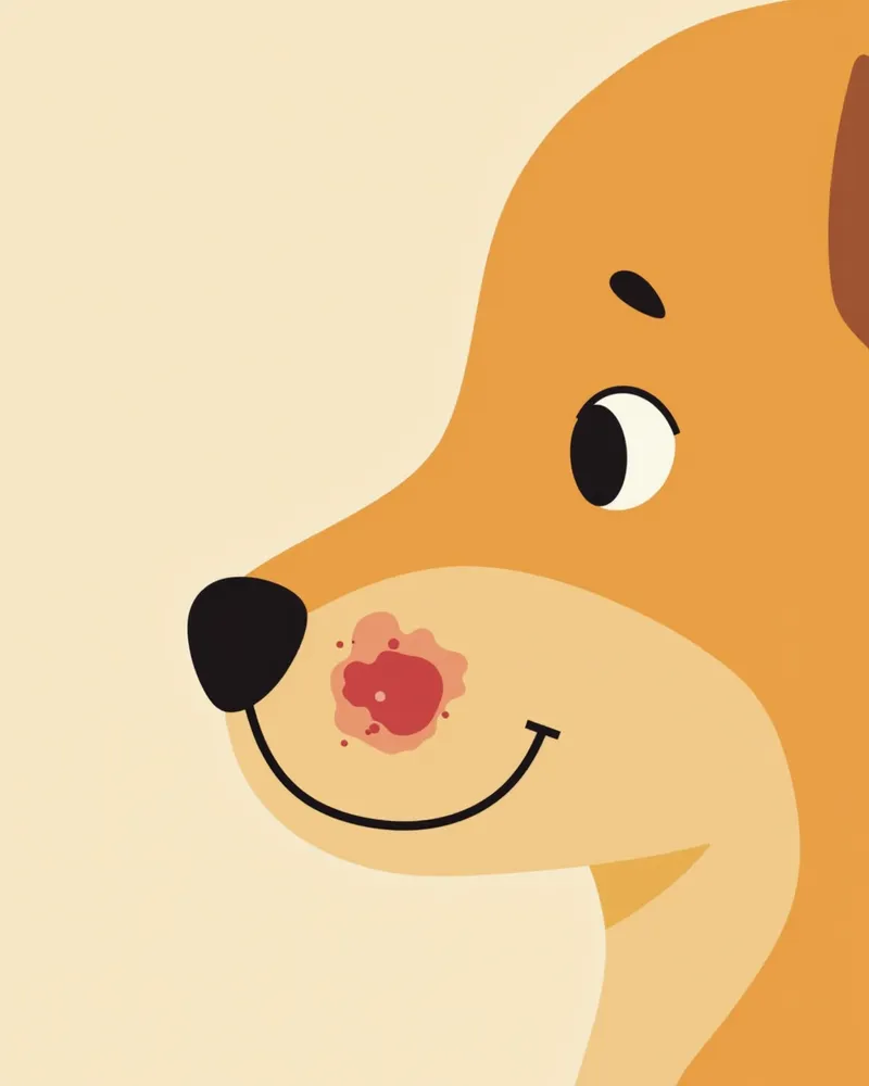
Ulcerative squamous cell carcinoma lesions on a dog's nose

Signs Requiring Immediate Veterinary Visit

If a lump persists for more than two weeks, or if you notice bleeding, pain, or an increase in size, consult your veterinarian immediately. Early diagnosis significantly improves treatment success rates. Extra caution is especially important for short-haired breeds.

How Is an Accurate Diagnosis Made?
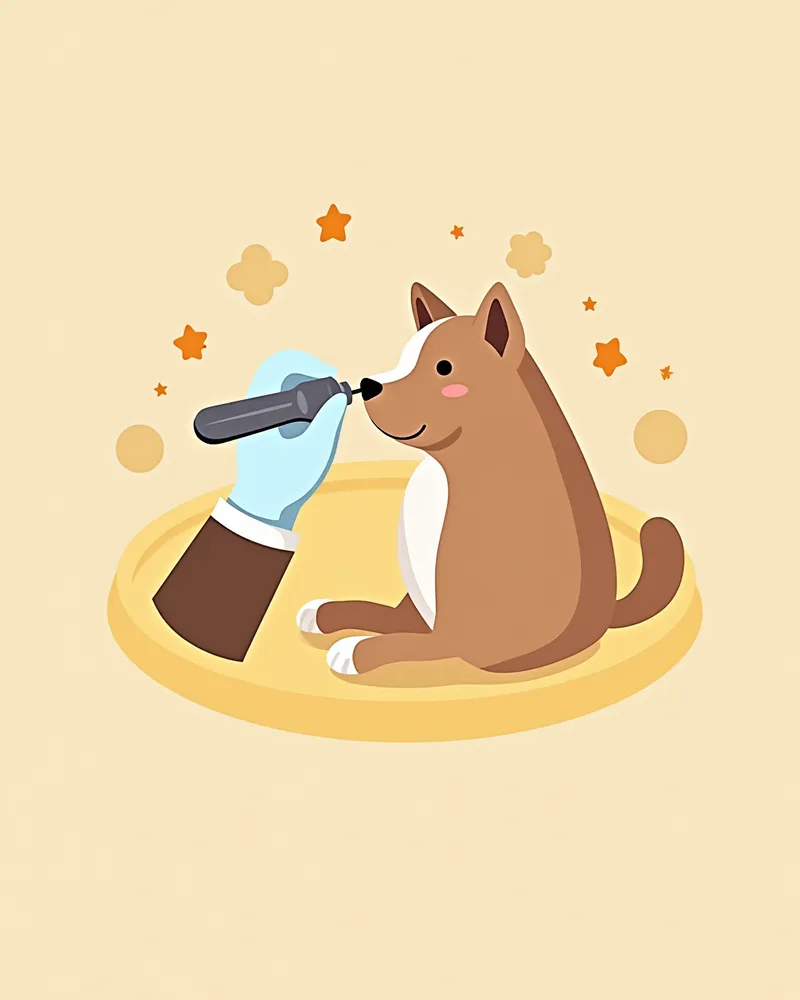
A tissue biopsy (histopathological examination) is essential for an accurate diagnosis. - Biopsy procedure: A representative tissue sample is taken from the lesion and examined under a microscope (histopathology) to determine whether it is malignant and, if so, its grade. - Examination method: Diagnosis is typically made by obtaining a representative tissue sample via incisional biopsy, with the specific method chosen based on clinical findings. - Metastasis assessment (staging): The presence of metastasis is evaluated through fine-needle aspiration of regional lymph nodes, three-view thoracic radiographs, and blood and serum tests. CT or MRI may be added when a detailed surgical plan is required. - Diagnostic accuracy: Histopathological examination is the most reliable method for definitive diagnosis. - Warning signs: If a lump persists for more than two weeks or changes in size or shape, immediate veterinary consultation is necessary.
A veterinarian performing a biopsy on a dog's skin.

What Are the Treatment Options?

Treatment options depend on the location, size, and extent of invasion of the lesion. Main approaches include wide surgical excision, cryosurgery, radiation therapy, and photodynamic therapy. Wide surgical excision is the primary treatment of choice; when the lesion is small, localized, and completely removed, a favorable prognosis can be expected. For advanced or invasive lesions, surgery may be combined with radiation or adjunctive therapies.
Surgical excision: The lesion is removed along with a sufficient margin of surrounding healthy tissue.
Cryosurgery: A practical option commonly used for small, superficial lesions.
Radiation therapy: Used as an alternative or adjunctive treatment for areas where complete surgical removal is difficult; these tumors show moderate radiosensitivity.
Photodynamic therapy: A treatment option applicable to early-stage, superficial lesions, such as those on the nasal planum.
A puppy undergoing radiation therapy

Comparison Table of Treatment Methods

ItemEffectSide effectsSuitable cases
Surgical excision (wide resection)HighSurgical pain, recovery period, recurrence with incomplete resectionWhen the lesion is small and localized
CryosurgeryModerate to highLocal irritation, temporary painSmall, superficial lesions
Radiation therapyModerate (moderate sensitivity)Dermatitis, hair lossSites where complete resection is difficult, adjuvant treatment
Photodynamic therapyModerate to highTemporary painEarly / superficial lesions such as the nose tip
ChemotherapyLimitedNausea, decreased appetiteNot recommended for localized SCC, role in metastatic disease not established

The treatment choice should be made after an accurate assessment by the veterinarian.

Precautions During Treatment

During treatment, it’s important to avoid touching or scratching the affected area. To reduce the risk of infection, limit contact with your pet and strictly follow your veterinarian’s instructions for medication and follow-up visits.

Reviewed by a veterinarian

Dr. Tony — Punnawat Phongkittirak

Dr. Tony — Punnawat Phongkittirak

Veterinarian

A veterinarian who majored in veterinary medicine at Khon Kaen University, Thailand, and completed the IVSA program at North Carolina State University in the United States. Drawing on clinical experience at animal hospitals, he works in the pet healthcare field and is dedicated to building a digital care environment that connects pet parents with veterinarians.

Frequently Asked Questions

Is canine squamous cell carcinoma treatable?
Yes, if the lesion is detected early and completely removed through surgery, there is a possibility of a cure. The success rate of treatment is high when there is no metastasis.
How can I prevent it?
Avoid going out during peak sun hours, and using sunscreen can also help. Extra care is especially important for breeds with short or thin coats.
How likely is it to recur?
The likelihood of recurrence after surgery depends on the depth of the lesion and whether it has metastasized. Regular check-ups and ongoing care are essential.
What should I do if my veterinarian doesn’t recommend surgery?
It is advisable to seek a second opinion at another veterinary clinic. Specialty hospitals can offer a range of treatment options beyond surgery, including radiation therapy and photodynamic therapy.
How much will the treatment cost?
Depending on the treatment—such as surgery, radiation therapy, or medication—costs can exceed $7,000. Since expenses vary significantly based on whether you have pet insurance and which veterinary clinic you choose, it’s a good idea to consult in advance.

Share

Related Guides

References

[1] Reimer, M.E. et al. (1999). Rectal ganglioneuroma in a dog. Journal of the American Animal Hospital Association, 35(2), 107–110.

[2] Hahn, K.A. et al. (2008). Masitinib is safe and effective for the treatment of canine mast cell tumors. Journal of Veterinary Internal Medicine, 22(6), 1301–1309.

[3] Small Animal Cytologic Diagnosis Canine and Feline Disease, 2nd Edition. (2012). Wiley-Blackwell.

This information is based on veterinary literature and does not replace diagnosis or treatment. Please consult a veterinarian for specific health concerns.

Metapet Co., Ltd. | CEO: Park Sung-yong | Business Reg. No. 417-88-02562 | Seoul, South Korea | Customer Center

What Is Canine Squamous Cell Carcinoma? Early Signs to Watch | Meongsiljang